Zhu Haiyan, Ai Yao, Zhang Jindi, Zhang Ji, Jin Juebin, Xie Congying, Su Huafang, Jin Xiance
Department of Gynecology, Shanghai First Maternal and Infant Hospital, Tongji University School of Medicine, Shanghai, China.
Department of Gynecology, The 1st Affiliated Hospital of Wenzhou Medical University, Wenzhou, China.
Front Oncol. 2021 Mar 25;11:642892. doi: 10.3389/fonc.2021.642892. eCollection 2021.
Non-invasive method to predict the histological subtypes preoperatively is essential for the overall management of ovarian cancer (OC). The feasibility of radiomics in the differentiating of epithelial ovarian cancer (EOC) and non-epithelial ovarian cancer (NEOC) based on computed tomography (CT) images was investigated.
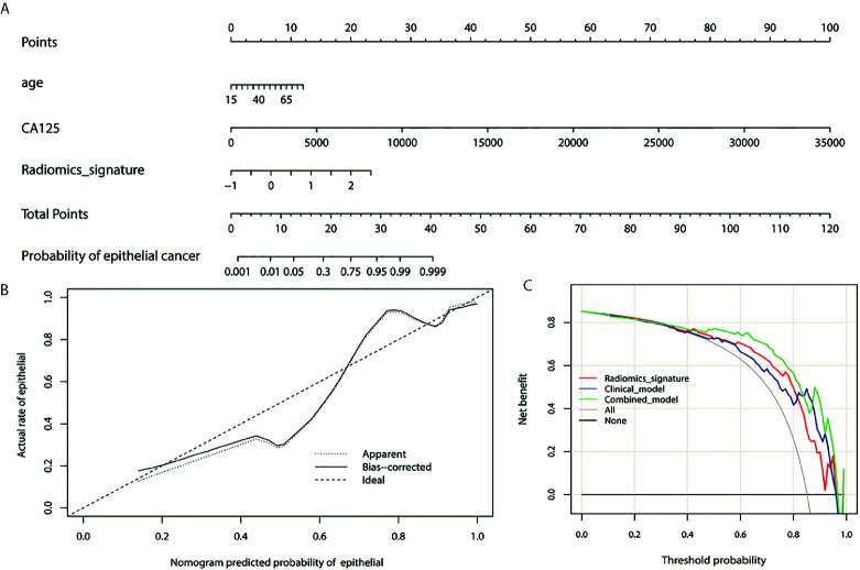
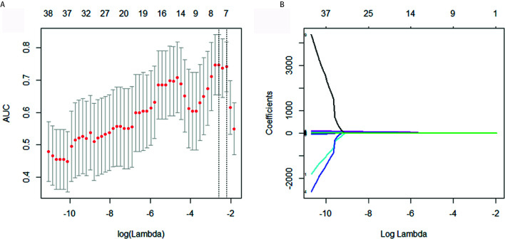
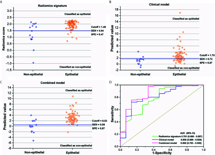
Radiomics features were extracted from preoperative CT for 101 patients with pathologically proven OC. Radiomics signature was built using the least absolute shrinkage and selection operator (LASSO) logistic regression. A nomogram was developed with the combination of radiomics features and clinical factors to differentiate EOC and NEOC.
Eight radiomics features were selected to build a radiomics signature with an area under curve (AUC) of 0.781 (95% confidence interval (CI), 0.666 -0.897) in the discrimination between EOC and NEOC. The AUC of the combined model integrating clinical factors and radiomics features was 0.869 (95% CI, 0.783 -0.955). The nomogram demonstrated that the combined model provides a better net benefit to predict histological subtypes compared with radiomics signature and clinical factors alone when the threshold probability is within a range from 0.43 to 0.97.
Nomogram developed with CT radiomics signature and clinical factors is feasible to predict the histological subtypes preoperative for patients with OC.
术前预测组织学亚型的非侵入性方法对于卵巢癌(OC)的整体管理至关重要。本研究探讨了基于计算机断层扫描(CT)图像的放射组学在鉴别上皮性卵巢癌(EOC)和非上皮性卵巢癌(NEOC)中的可行性。
从101例经病理证实的OC患者的术前CT图像中提取放射组学特征。使用最小绝对收缩和选择算子(LASSO)逻辑回归构建放射组学特征。将放射组学特征与临床因素相结合,开发出一种列线图,用于鉴别EOC和NEOC。
选择了8个放射组学特征来构建放射组学特征,在区分EOC和NEOC时,曲线下面积(AUC)为0.781(95%置信区间(CI),0.666 - 0.897)。整合临床因素和放射组学特征的联合模型的AUC为0.869(95% CI,0.783 - 0.955)。列线图显示,当阈值概率在0.43至0.97范围内时,与单独的放射组学特征和临床因素相比,联合模型在预测组织学亚型方面提供了更好的净效益。
利用CT放射组学特征和临床因素开发的列线图对于术前预测OC患者的组织学亚型是可行的。