• 文献检索
  • 文档翻译
  • 深度研究
  • 学术资讯
  • Suppr Zotero 插件Zotero 插件
  • 邀请有礼
  • 套餐&价格
  • 历史记录
应用&插件
Suppr Zotero 插件Zotero 插件浏览器插件Mac 客户端Windows 客户端微信小程序
定价
高级版会员购买积分包购买API积分包
服务
文献检索文档翻译深度研究API 文档MCP 服务
关于我们
关于 Suppr公司介绍联系我们用户协议隐私条款
关注我们

Suppr 超能文献

核心技术专利:CN118964589B侵权必究
粤ICP备2023148730 号-1Suppr @ 2026

文献检索

告别复杂PubMed语法,用中文像聊天一样搜索,搜遍4000万医学文献。AI智能推荐,让科研检索更轻松。

立即免费搜索

文件翻译

保留排版,准确专业,支持PDF/Word/PPT等文件格式,支持 12+语言互译。

免费翻译文档

深度研究

AI帮你快速写综述,25分钟生成高质量综述,智能提取关键信息,辅助科研写作。

立即免费体验

干细胞驱动的血吸虫发育的单细胞解构

Single-cell deconstruction of stem-cell-driven schistosome development.

作者信息

Nanes Sarfati Dania, Li Pengyang, Tarashansky Alexander J, Wang Bo

机构信息

Department of Biology, Stanford University, Stanford, CA, USA.

Department of Bioengineering, Stanford University, Stanford, CA, USA.

出版信息

Trends Parasitol. 2021 Sep;37(9):790-802. doi: 10.1016/j.pt.2021.03.005. Epub 2021 Apr 20.

DOI:10.1016/j.pt.2021.03.005
PMID:33893056
原文链接:https://pmc.ncbi.nlm.nih.gov/articles/PMC8364473/
Abstract

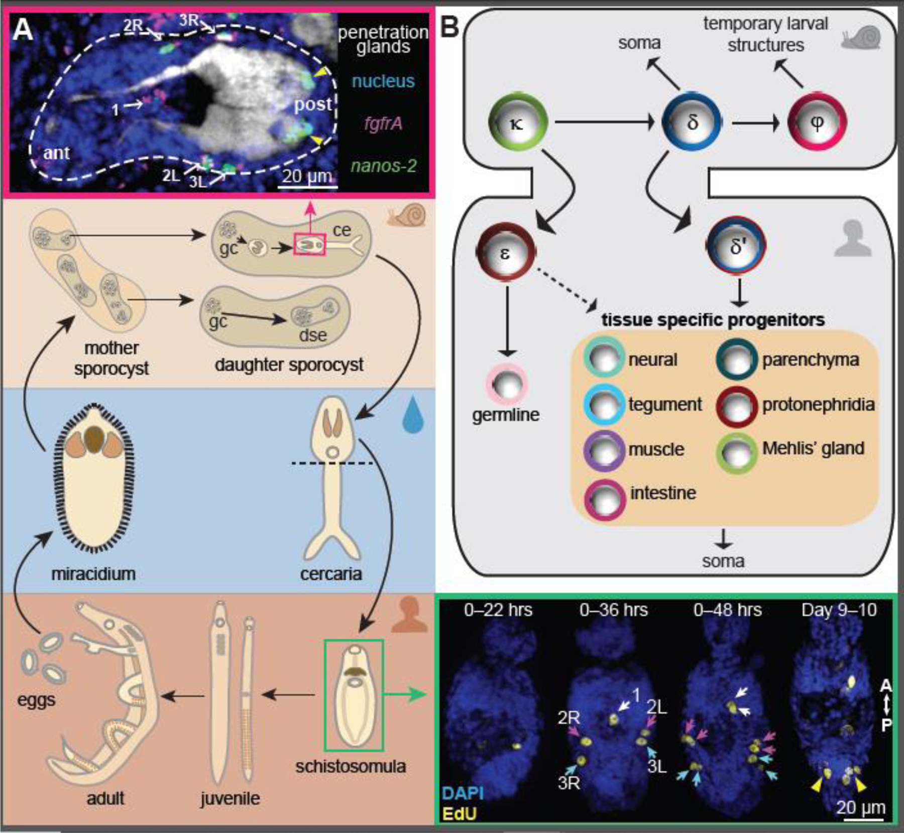
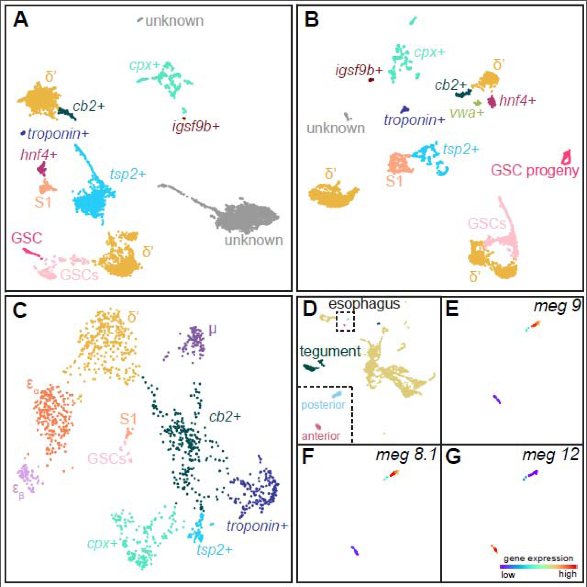
Schistosomes cause one of the most devastating neglected tropical diseases, schistosomiasis. Their transmission is accomplished through a complex life cycle with two obligate hosts and requires multiple radically different body plans specialized for infecting and reproducing in each host. Recent single-cell transcriptomic studies on several schistosome body plans provide a comprehensive map of their cell types, which include stem cells and their differentiated progeny along an intricate developmental hierarchy. This progress not only extends our understanding of the basic biology of the schistosome life cycle but can also inform new therapeutic and preventive strategies against the disease, as blocking the development of specific cell types through genetic manipulations has shown promise in inhibiting parasite survival, growth, and reproduction.

摘要

血吸虫引发了最具破坏性的被忽视热带病之一——血吸虫病。它们通过一个涉及两个必需宿主的复杂生命周期来完成传播,并且需要多个截然不同的身体结构,这些结构专门用于在每个宿主中进行感染和繁殖。最近针对几种血吸虫身体结构的单细胞转录组学研究提供了其细胞类型的全面图谱,其中包括干细胞及其沿着复杂发育层级分化的后代。这一进展不仅扩展了我们对血吸虫生命周期基本生物学的理解,还能为针对该疾病的新治疗和预防策略提供依据,因为通过基因操作阻断特定细胞类型的发育已显示出抑制寄生虫存活、生长和繁殖的潜力。

https://cdn.ncbi.nlm.nih.gov/pmc/blobs/2ca7/8364473/4d88dafa0f4d/nihms-1696722-f0002.jpg
https://cdn.ncbi.nlm.nih.gov/pmc/blobs/2ca7/8364473/7ec33a6bfea4/nihms-1696722-f0001.jpg
https://cdn.ncbi.nlm.nih.gov/pmc/blobs/2ca7/8364473/4d88dafa0f4d/nihms-1696722-f0002.jpg
https://cdn.ncbi.nlm.nih.gov/pmc/blobs/2ca7/8364473/7ec33a6bfea4/nihms-1696722-f0001.jpg
https://cdn.ncbi.nlm.nih.gov/pmc/blobs/2ca7/8364473/4d88dafa0f4d/nihms-1696722-f0002.jpg

相似文献

1
Single-cell deconstruction of stem-cell-driven schistosome development.干细胞驱动的血吸虫发育的单细胞解构
Trends Parasitol. 2021 Sep;37(9):790-802. doi: 10.1016/j.pt.2021.03.005. Epub 2021 Apr 20.
2
The Tao survivorship of schistosomes: implications for schistosomiasis control.血吸虫的生存之道:对血吸虫病控制的启示
Int J Parasitol. 2016 Jun;46(7):453-63. doi: 10.1016/j.ijpara.2016.01.002. Epub 2016 Feb 9.
3
Schistosomiasis as a disease of stem cells.血吸虫病作为一种干细胞疾病。
Curr Opin Genet Dev. 2016 Oct;40:95-102. doi: 10.1016/j.gde.2016.06.010. Epub 2016 Jul 5.
4
Stem cell heterogeneity drives the parasitic life cycle of .干细胞异质性驱动 的寄生生活史。
Elife. 2018 Jul 10;7:e35449. doi: 10.7554/eLife.35449.
5
Applications for profiling the schistosome transcriptome.用于对血吸虫转录组进行分析的应用。
Trends Parasitol. 2010 Sep;26(9):434-9. doi: 10.1016/j.pt.2010.04.009. Epub 2010 May 31.
6
Innovations and Advances in Schistosome Stem Cell Research.血吸虫干细胞研究的创新与进展。
Front Immunol. 2021 Mar 5;12:599014. doi: 10.3389/fimmu.2021.599014. eCollection 2021.
7
Schistosoma genomics: new perspectives on schistosome biology and host-parasite interaction.血吸虫基因组学:血吸虫生物学及宿主-寄生虫相互作用的新视角
Annu Rev Genomics Hum Genet. 2009;10:211-40. doi: 10.1146/annurev-genom-082908-150036.
8
Single-sex schistosomiasis: a mini review.单性血吸虫病:小型综述。
Front Immunol. 2023 Apr 19;14:1158805. doi: 10.3389/fimmu.2023.1158805. eCollection 2023.
9
Planarians to schistosomes: an overview of flatworm cell-types and regulators.从涡虫到血吸虫:扁形动物细胞类型和调控因子概述。
J Helminthol. 2023 Jan 16;97:e7. doi: 10.1017/S0022149X22000621.
10
The use of imaging to detect schistosomes and diagnose schistosomiasis.利用影像学手段检测并诊断血吸虫病。
Parasite Immunol. 2013 Sep-Oct;35(9-10):295-301. doi: 10.1111/pim.12040.

引用本文的文献

1
Abundant genetic variation is retained in many laboratory schistosome populations.许多实验室血吸虫种群中保留了丰富的遗传变异。
PLoS Pathog. 2025 Aug 20;21(8):e1013439. doi: 10.1371/journal.ppat.1013439. eCollection 2025 Aug.
2
The retinoic acid family-like nuclear receptor SmRAR identified by single-cell transcriptomics of ovarian cells controls oocyte differentiation in Schistosoma mansoni.通过对卵巢细胞进行单细胞转录组学鉴定出的类视黄酸家族核受体SmRAR控制曼氏血吸虫的卵母细胞分化。
Nucleic Acids Res. 2025 Feb 8;53(4). doi: 10.1093/nar/gkae1228.
3
Schistosome esophageal gland factor MEG-8.2 drives host cell lysis and interacts with host immune proteins.

本文引用的文献

1
Mapping single-cell atlases throughout Metazoa unravels cell type evolution.绘制整个后生动物单细胞图谱揭示了细胞类型的进化。
Elife. 2021 May 4;10:e66747. doi: 10.7554/eLife.66747.
2
Single-cell analysis of Schistosoma mansoni identifies a conserved genetic program controlling germline stem cell fate.单细胞分析曼氏血吸虫鉴定出一个保守的遗传程序,控制生殖干细胞命运。
Nat Commun. 2021 Jan 20;12(1):485. doi: 10.1038/s41467-020-20794-w.
3
Single-cell atlas of the first intra-mammalian developmental stage of the human parasite Schistosoma mansoni.
血吸虫食管腺因子MEG-8.2驱动宿主细胞裂解并与宿主免疫蛋白相互作用。
bioRxiv. 2024 Nov 15:2024.11.15.623777. doi: 10.1101/2024.11.15.623777.
4
Abundant genetic variation is retained in many laboratory schistosome populations.许多实验室血吸虫种群中保留了丰富的遗传变异。
bioRxiv. 2024 Oct 24:2024.10.21.619418. doi: 10.1101/2024.10.21.619418.
5
Flexible use of conserved motif vocabularies constrains genome access in cell type evolution.保守基序词汇的灵活运用在细胞类型进化中限制了基因组的可及性。
bioRxiv. 2024 Sep 6:2024.09.03.611027. doi: 10.1101/2024.09.03.611027.
6
A single-cell atlas of the miracidium larva of reveals cell types, developmental pathways, and tissue architecture.揭示尾蚴幼虫细胞类型、发育途径和组织架构的单细胞图谱。
Elife. 2024 Aug 27;13:RP95628. doi: 10.7554/eLife.95628.
7
Trematode Genomics and Proteomics.吸虫组学和蛋白质组学。
Adv Exp Med Biol. 2024;1454:507-539. doi: 10.1007/978-3-031-60121-7_13.
8
Single-cell transcriptomics of the human parasite Schistosoma mansoni first intra-molluscan stage reveals tentative tegumental and stem-cell regulators.人类寄生虫曼氏血吸虫首个软体动物内阶段的单细胞转录组学揭示了初步的表皮和干细胞调节因子。
Sci Rep. 2024 Mar 12;14(1):5974. doi: 10.1038/s41598-024-55790-3.
9
Functional genomics in Spiralia.环节动物的功能基因组学。
Brief Funct Genomics. 2023 Nov 17;22(6):487-497. doi: 10.1093/bfgp/elad036.
10
CRISPR interference for sequence-specific regulation of fibroblast growth factor receptor A in .CRISPR 干扰在. 中对成纤维细胞生长因子受体 A 的序列特异性调控
Front Immunol. 2023 Jan 13;13:1105719. doi: 10.3389/fimmu.2022.1105719. eCollection 2022.
人类寄生虫曼氏血吸虫在哺乳动物体内第一个发育阶段的单细胞图谱。
Nat Commun. 2020 Dec 18;11(1):6411. doi: 10.1038/s41467-020-20092-5.
4
A single-cell RNA-seq atlas of identifies a key regulator of blood feeding.一份关于[具体内容缺失]的单细胞RNA测序图谱鉴定出了吸血的关键调节因子。
Science. 2020 Sep 25;369(6511):1644-1649. doi: 10.1126/science.abb7709.
5
The esophageal gland mediates host immune evasion by the human parasite .食管腺介导人体寄生虫的宿主免疫逃避。
Proc Natl Acad Sci U S A. 2020 Aug 11;117(32):19299-19309. doi: 10.1073/pnas.2006553117. Epub 2020 Jul 31.
6
Schistosome migration in the definitive host.血吸虫在终宿主中的迁移。
PLoS Negl Trop Dis. 2020 Apr 2;14(4):e0007951. doi: 10.1371/journal.pntd.0007951. eCollection 2020 Apr.
7
Concomitant Immunity and Worm Senescence May Drive Schistosomiasis Epidemiological Patterns: An Eco-Evolutionary Perspective.伴随免疫和蠕虫衰老可能驱动血吸虫病流行病学模式:生态进化观点。
Front Immunol. 2020 Feb 25;11:160. doi: 10.3389/fimmu.2020.00160. eCollection 2020.
8
Self-assembling manifolds in single-cell RNA sequencing data.单细胞 RNA 测序数据中的自组装流形。
Elife. 2019 Sep 16;8:e48994. doi: 10.7554/eLife.48994.
9
Systematically improved in vitro culture conditions reveal new insights into the reproductive biology of the human parasite Schistosoma mansoni.系统地改进体外培养条件揭示了人类寄生虫曼氏血吸虫生殖生物学的新见解。
PLoS Biol. 2019 May 8;17(5):e3000254. doi: 10.1371/journal.pbio.3000254. eCollection 2019 May.
10
The Cellular and Molecular Basis for Planarian Regeneration.涡虫再生的细胞和分子基础。
Cell. 2018 Oct 4;175(2):327-345. doi: 10.1016/j.cell.2018.09.021.