Department of Imaging Physics, Delft University of Technology, Delft, The Netherlands.
Department of NanoBiophotonics, Max Planck Institute for Biophysical Chemistry, Göttingen, Germany.
Nat Commun. 2021 May 14;12(1):2847. doi: 10.1038/s41467-021-22006-5.
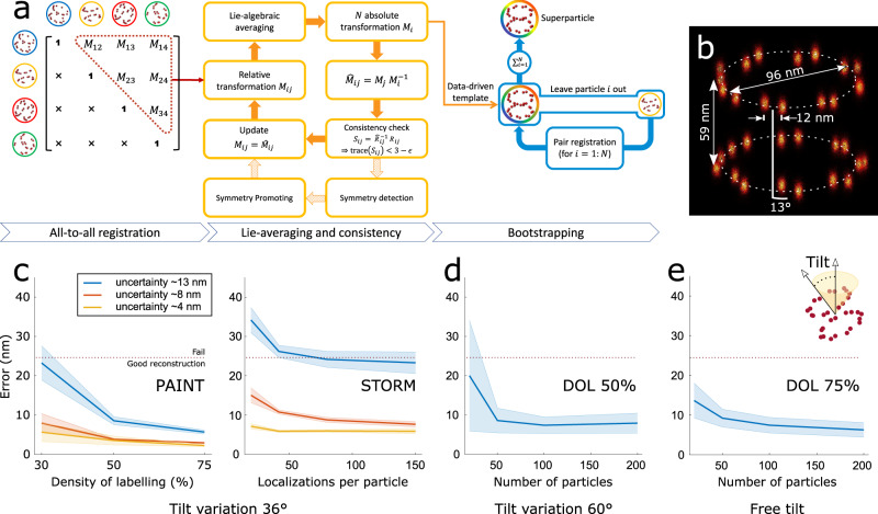
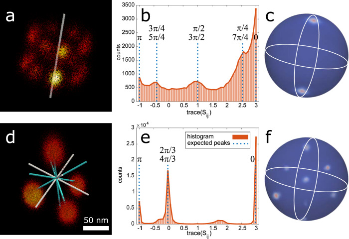
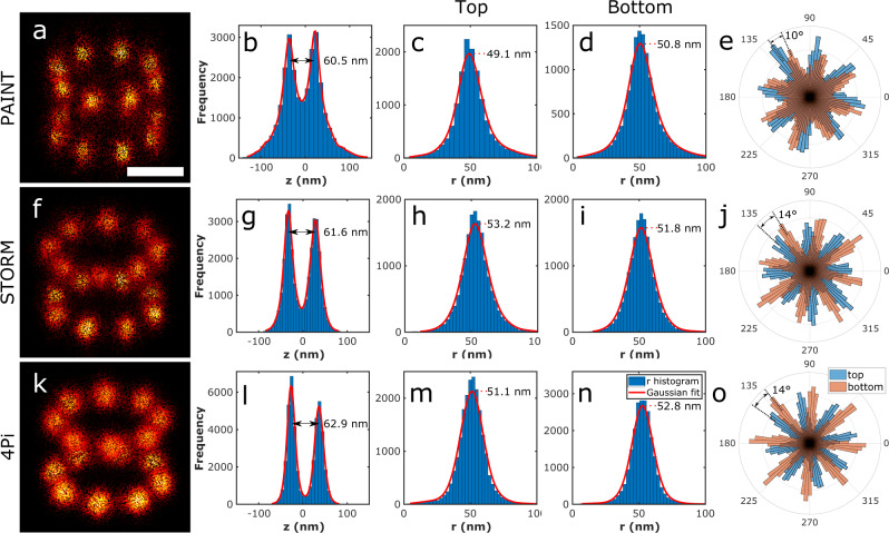
Single molecule localization microscopy offers in principle resolution down to the molecular level, but in practice this is limited primarily by incomplete fluorescent labeling of the structure. This missing information can be completed by merging information from many structurally identical particles. In this work, we present an approach for 3D single particle analysis in localization microscopy which hugely increases signal-to-noise ratio and resolution and enables determining the symmetry groups of macromolecular complexes. Our method does not require a structural template, and handles anisotropic localization uncertainties. We demonstrate 3D reconstructions of DNA-origami tetrahedrons, Nup96 and Nup107 subcomplexes of the nuclear pore complex acquired using multiple single molecule localization microscopy techniques, with their structural symmetry deducted from the data.
单分子定位显微镜原则上可以提供达到分子水平的分辨率,但实际上这主要受到结构中不完全荧光标记的限制。可以通过合并来自许多结构相同的粒子的信息来完成这些缺失的信息。在这项工作中,我们提出了一种用于定位显微镜中单粒子分析的方法,该方法大大提高了信噪比和分辨率,并能够确定大分子复合物的对称群。我们的方法不需要结构模板,并处理各向异性的定位不确定性。我们展示了使用多种单分子定位显微镜技术获得的 DNA 折纸四面体、核孔复合物的 Nup96 和 Nup107 亚复合物的 3D 重建,其结构对称性是从数据中推断出来的。