RENCI, University of North Carolina at Chapel Hill, 100 Europa Drive, Suite 540, Chapel Hill, NC, 27517, USA.
UNC Neuroscience Center, University of North Carolina at Chapel Hill, 116 Manning Drive, CB# 7250, Chapel Hill, NC, 27599, USA.
BMC Bioinformatics. 2021 May 22;22(1):260. doi: 10.1186/s12859-021-04202-8.
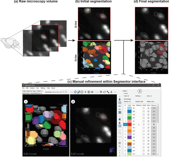
Recent advances in tissue clearing techniques, combined with high-speed image acquisition through light sheet microscopy, enable rapid three-dimensional (3D) imaging of biological specimens, such as whole mouse brains, in a matter of hours. Quantitative analysis of such 3D images can help us understand how changes in brain structure lead to differences in behavior or cognition, but distinguishing densely packed features of interest, such as nuclei, from background can be challenging. Recent deep learning-based nuclear segmentation algorithms show great promise for automated segmentation, but require large numbers of accurate manually labeled nuclei as training data.
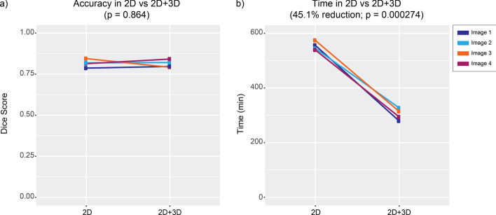
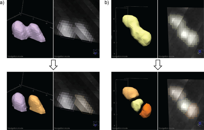
We present Segmentor, an open-source tool for reliable, efficient, and user-friendly manual annotation and refinement of objects (e.g., nuclei) within 3D light sheet microscopy images. Segmentor employs a hybrid 2D-3D approach for visualizing and segmenting objects and contains features for automatic region splitting, designed specifically for streamlining the process of 3D segmentation of nuclei. We show that editing simultaneously in 2D and 3D using Segmentor significantly decreases time spent on manual annotations without affecting accuracy as compared to editing the same set of images with only 2D capabilities.
Segmentor is a tool for increased efficiency of manual annotation and refinement of 3D objects that can be used to train deep learning segmentation algorithms, and is available at https://www.nucleininja.org/ and https://github.com/RENCI/Segmentor .
组织透明化技术的最新进展,结合光片显微镜的高速图像采集,使得对生物标本(如整个老鼠大脑)进行快速三维(3D)成像成为可能,整个过程只需几个小时。对这些 3D 图像进行定量分析可以帮助我们了解大脑结构的变化如何导致行为或认知的差异,但从背景中区分出诸如细胞核等密集排列的感兴趣特征可能具有挑战性。最近基于深度学习的核分割算法在自动分割方面显示出巨大的潜力,但需要大量准确的手动标记核作为训练数据。
我们提出了 Segmentor,这是一种用于可靠、高效和用户友好的 3D 光片显微镜图像中手动注释和细化对象(如细胞核)的开源工具。Segmentor 采用了 2D-3D 混合方法来可视化和分割对象,并包含了用于自动区域分割的功能,专门用于简化细胞核的 3D 分割过程。我们表明,与仅使用 2D 功能编辑相同的一组图像相比,使用 Segmentor 在 2D 和 3D 中同时编辑可以显著减少手动注释所需的时间,而不会影响准确性。
Segmentor 是一种提高手动注释和细化 3D 对象效率的工具,可用于训练深度学习分割算法,可在 https://www.nucleininja.org/ 和 https://github.com/RENCI/Segmentor 上获得。