• 文献检索
  • 文档翻译
  • 深度研究
  • 学术资讯
  • Suppr Zotero 插件Zotero 插件
  • 邀请有礼
  • 套餐&价格
  • 历史记录
应用&插件
Suppr Zotero 插件Zotero 插件浏览器插件Mac 客户端Windows 客户端微信小程序
定价
高级版会员购买积分包购买API积分包
服务
文献检索文档翻译深度研究API 文档MCP 服务
关于我们
关于 Suppr公司介绍联系我们用户协议隐私条款
关注我们

Suppr 超能文献

核心技术专利:CN118964589B侵权必究
粤ICP备2023148730 号-1Suppr @ 2026

文献检索

告别复杂PubMed语法,用中文像聊天一样搜索,搜遍4000万医学文献。AI智能推荐,让科研检索更轻松。

立即免费搜索

文件翻译

保留排版,准确专业,支持PDF/Word/PPT等文件格式,支持 12+语言互译。

免费翻译文档

深度研究

AI帮你快速写综述,25分钟生成高质量综述,智能提取关键信息,辅助科研写作。

立即免费体验

基于深度学习的人体胸部计算机断层扫描异常检测与智能严重程度评估:以SARS-CoV-2评估为例

Abnormality detection and intelligent severity assessment of human chest computed tomography scans using deep learning: a case study on SARS-COV-2 assessment.

作者信息

Ibrahim Mohamed Ramzy, Youssef Sherin M, Fathalla Karma M

机构信息

Computer Engineering Department, Arab Academy for Science, Technology and Maritime Transport (AASTMT), Alexandria, 1029 Egypt.

出版信息

J Ambient Intell Humaniz Comput. 2023;14(5):5665-5688. doi: 10.1007/s12652-021-03282-x. Epub 2021 May 25.

DOI:10.1007/s12652-021-03282-x
PMID:34055098
原文链接:https://pmc.ncbi.nlm.nih.gov/articles/PMC8147594/
Abstract

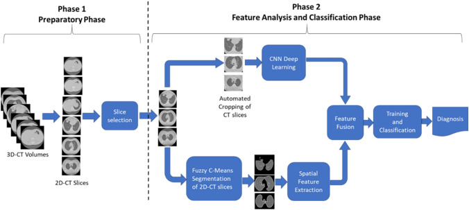
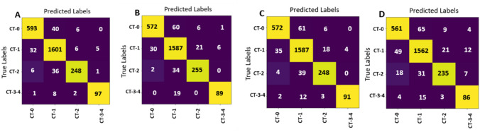
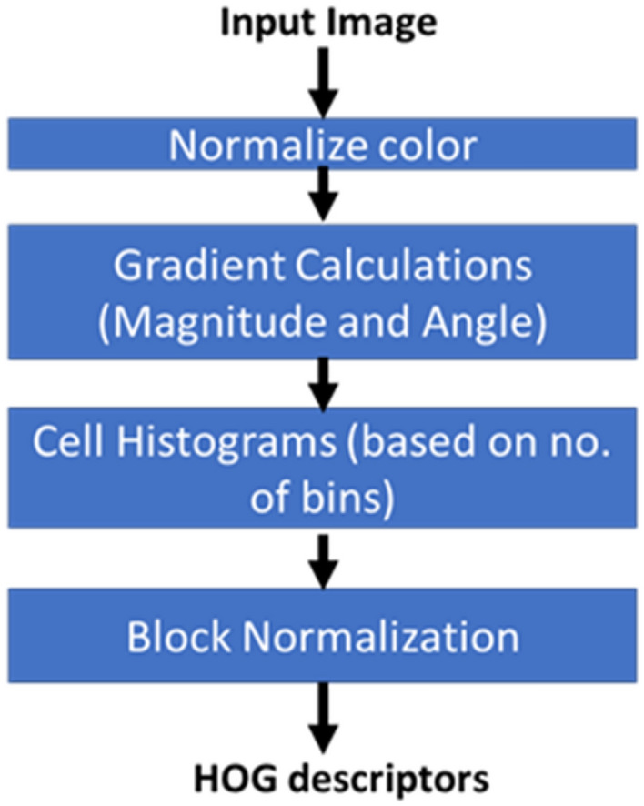
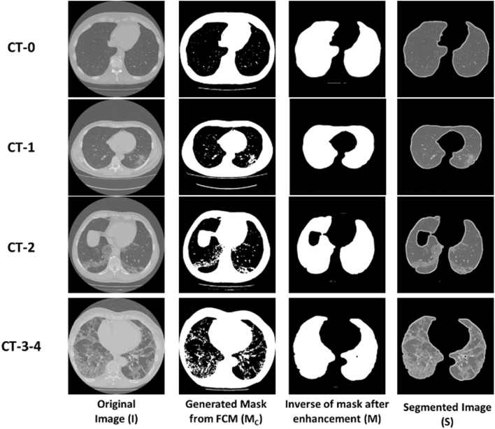
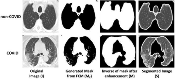
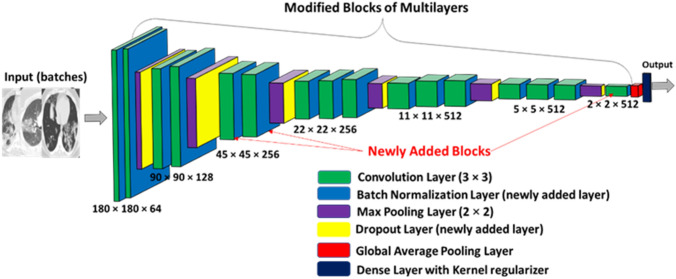
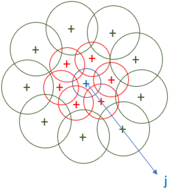
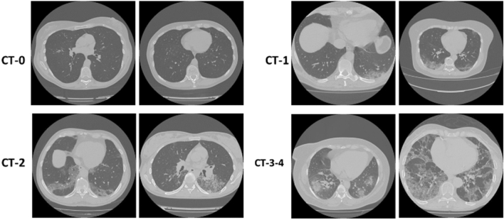
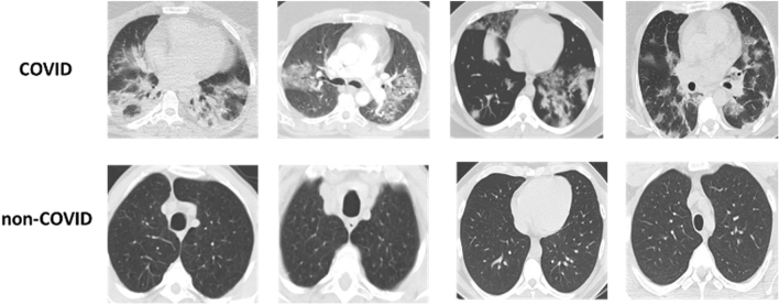
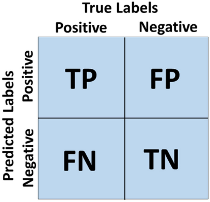
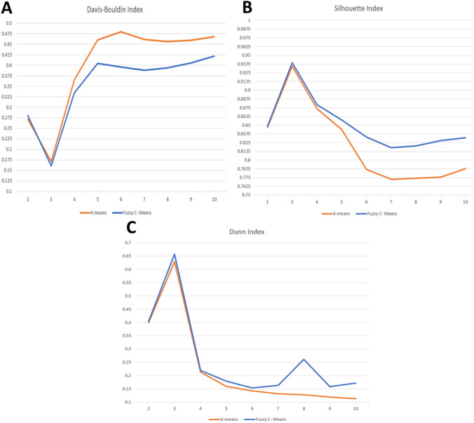
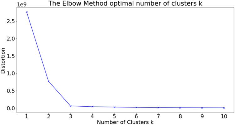
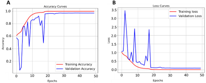
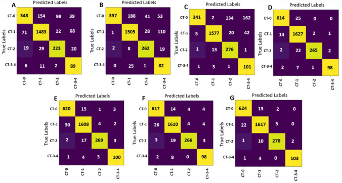
Different respiratory infections cause abnormal symptoms in lung parenchyma that show in chest computed tomography. Since December 2019, the SARS-COV-2 virus, which is the causative agent of COVID-19, has invaded the world causing high numbers of infections and deaths. The infection with SARS-COV-2 virus shows an abnormality in lung parenchyma that can be effectively detected using Computed Tomography (CT) imaging. In this paper, a novel computer aided framework (COV-CAF) is proposed for classifying the severity degree of the infection from 3D Chest Volumes. COV-CAF fuses traditional and deep learning approaches. The proposed COV-CAF consists of two phases: the preparatory phase and the feature analysis and classification phase. The preparatory phase handles 3D-CT volumes and presents an effective cut choice strategy for choosing informative CT slices. The feature analysis and classification phase incorporate fuzzy clustering for automatic Region of Interest (RoI) segmentation and feature fusion. In feature fusion, automatic features are extracted from a newly introduced Convolution Neural Network (Norm-VGG16) and are fused with spatial hand-crafted features extracted from segmented RoI. Experiments are conducted on MosMedData: Chest CT Scans with COVID-19 Related Findings with COVID-19 severity classes and SARS-COV-2 CT-Scan benchmark datasets. The proposed COV-CAF achieved remarkable results on both datasets. On MosMedData dataset, it achieved an overall accuracy of 97.76% and average sensitivity of 96.73%, while on SARS-COV-2 CT-Scan dataset it achieves an overall accuracy and sensitivity 97.59% and 98.41% respectively.

摘要

不同的呼吸道感染会导致肺实质出现异常症状,这些症状会在胸部计算机断层扫描中显示出来。自2019年12月以来,导致COVID-19的病原体SARS-CoV-2病毒侵袭全球,造成大量感染和死亡。感染SARS-CoV-2病毒会导致肺实质出现异常,使用计算机断层扫描(CT)成像可以有效检测到这种异常。本文提出了一种新颖的计算机辅助框架(COV-CAF),用于从3D胸部容积中对感染的严重程度进行分类。COV-CAF融合了传统方法和深度学习方法。所提出的COV-CAF包括两个阶段:准备阶段和特征分析与分类阶段。准备阶段处理3D-CT容积,并提出一种有效的切片选择策略,用于选择信息丰富的CT切片。特征分析与分类阶段结合模糊聚类进行自动感兴趣区域(RoI)分割和特征融合。在特征融合中,从新引入的卷积神经网络(Norm-VGG16)中提取自动特征,并与从分割后的RoI中提取的空间手工特征进行融合。在MosMedData数据集上进行了实验:该数据集包含与COVID-19相关发现的胸部CT扫描以及COVID-19严重程度分类和SARS-CoV-2 CT扫描基准数据集。所提出的COV-CAF在两个数据集上均取得了显著成果。在MosMedData数据集上,其总体准确率达到97.76%,平均灵敏度为96.73%,而在SARS-CoV-2 CT扫描数据集上,其总体准确率和灵敏度分别达到97.59%和98.41%。

https://cdn.ncbi.nlm.nih.gov/pmc/blobs/5406/8147594/44acce2c58b8/12652_2021_3282_Fig16_HTML.jpg
https://cdn.ncbi.nlm.nih.gov/pmc/blobs/5406/8147594/e547cd9793ff/12652_2021_3282_Fig1_HTML.jpg
https://cdn.ncbi.nlm.nih.gov/pmc/blobs/5406/8147594/78fa12ae1d94/12652_2021_3282_Fig2_HTML.jpg
https://cdn.ncbi.nlm.nih.gov/pmc/blobs/5406/8147594/687464a21522/12652_2021_3282_Fig3_HTML.jpg
https://cdn.ncbi.nlm.nih.gov/pmc/blobs/5406/8147594/f498dc1eba3c/12652_2021_3282_Fig4_HTML.jpg
https://cdn.ncbi.nlm.nih.gov/pmc/blobs/5406/8147594/8a5bb830221b/12652_2021_3282_Fig5_HTML.jpg
https://cdn.ncbi.nlm.nih.gov/pmc/blobs/5406/8147594/b7454cec5bf8/12652_2021_3282_Fig6_HTML.jpg
https://cdn.ncbi.nlm.nih.gov/pmc/blobs/5406/8147594/014c66781b27/12652_2021_3282_Fig7_HTML.jpg
https://cdn.ncbi.nlm.nih.gov/pmc/blobs/5406/8147594/f6c3e0e2fe40/12652_2021_3282_Fig8_HTML.jpg
https://cdn.ncbi.nlm.nih.gov/pmc/blobs/5406/8147594/2cd1219742dc/12652_2021_3282_Fig9_HTML.jpg
https://cdn.ncbi.nlm.nih.gov/pmc/blobs/5406/8147594/418fcaea7f53/12652_2021_3282_Fig10_HTML.jpg
https://cdn.ncbi.nlm.nih.gov/pmc/blobs/5406/8147594/6e053fe3aa4c/12652_2021_3282_Fig11_HTML.jpg
https://cdn.ncbi.nlm.nih.gov/pmc/blobs/5406/8147594/22c64ae413c9/12652_2021_3282_Fig12_HTML.jpg
https://cdn.ncbi.nlm.nih.gov/pmc/blobs/5406/8147594/0a128d04c7a0/12652_2021_3282_Fig13_HTML.jpg
https://cdn.ncbi.nlm.nih.gov/pmc/blobs/5406/8147594/e95f2c9a82f8/12652_2021_3282_Fig14_HTML.jpg
https://cdn.ncbi.nlm.nih.gov/pmc/blobs/5406/8147594/e2766ad60851/12652_2021_3282_Fig15_HTML.jpg
https://cdn.ncbi.nlm.nih.gov/pmc/blobs/5406/8147594/44acce2c58b8/12652_2021_3282_Fig16_HTML.jpg

相似文献

1
Abnormality detection and intelligent severity assessment of human chest computed tomography scans using deep learning: a case study on SARS-COV-2 assessment.基于深度学习的人体胸部计算机断层扫描异常检测与智能严重程度评估:以SARS-CoV-2评估为例
J Ambient Intell Humaniz Comput. 2023;14(5):5665-5688. doi: 10.1007/s12652-021-03282-x. Epub 2021 May 25.
2
An intelligence design for detection and classification of COVID19 using fusion of classical and convolutional neural network and improved microscopic features selection approach.基于经典与卷积神经网络融合及改进的微观特征选择方法的 COVID19 检测与分类智能设计。
Microsc Res Tech. 2021 Oct;84(10):2254-2267. doi: 10.1002/jemt.23779. Epub 2021 May 8.
3
An automated diagnosis and classification of COVID-19 from chest CT images using a transfer learning-based convolutional neural network.利用基于迁移学习的卷积神经网络对 chest CT 图像进行 COVID-19 的自动诊断和分类。
Comput Biol Med. 2022 May;144:105383. doi: 10.1016/j.compbiomed.2022.105383. Epub 2022 Mar 10.
4
A CNN-LSTM network with multi-level feature extraction-based approach for automated detection of coronavirus from CT scan and X-ray images.一种基于多级特征提取的卷积神经网络-长短期记忆网络,用于从CT扫描和X光图像中自动检测冠状病毒。
Appl Soft Comput. 2021 Dec;113:107918. doi: 10.1016/j.asoc.2021.107918. Epub 2021 Sep 27.
5
COVID-DSNet: A novel deep convolutional neural network for detection of coronavirus (SARS-CoV-2) cases from CT and Chest X-Ray images.COVID-DSNet:一种新型深度卷积神经网络,用于从 CT 和胸部 X 光图像中检测冠状病毒(SARS-CoV-2)病例。
Artif Intell Med. 2022 Dec;134:102427. doi: 10.1016/j.artmed.2022.102427. Epub 2022 Oct 17.
6
A wavelet-based deep learning pipeline for efficient COVID-19 diagnosis via CT slices.一种基于小波的深度学习管道,用于通过CT切片高效诊断新冠肺炎。
Appl Soft Comput. 2022 Oct;128:109401. doi: 10.1016/j.asoc.2022.109401. Epub 2022 Jul 29.
7
COVID-19 detection from lung CT-Scans using a fuzzy integral-based CNN ensemble.基于模糊积分的卷积神经网络集成从肺部CT扫描中检测新型冠状病毒肺炎
Comput Biol Med. 2021 Nov;138:104895. doi: 10.1016/j.compbiomed.2021.104895. Epub 2021 Oct 1.
8
A novel adaptive cubic quasi-Newton optimizer for deep learning based medical image analysis tasks, validated on detection of COVID-19 and segmentation for COVID-19 lung infection, liver tumor, and optic disc/cup.一种用于深度学习的新型自适应三次拟牛顿优化器,在 COVID-19 检测和 COVID-19 肺部感染、肝脏肿瘤以及视盘/杯分割等医学图像分析任务中得到验证。
Med Phys. 2023 Mar;50(3):1528-1538. doi: 10.1002/mp.15969. Epub 2022 Oct 6.
9
Computed tomography-based COVID-19 triage through a deep neural network using mask-weighted global average pooling.基于计算机断层扫描的 COVID-19 分诊的深度神经网络方法,使用口罩加权全局平均池化。
Front Cell Infect Microbiol. 2023 Mar 3;13:1116285. doi: 10.3389/fcimb.2023.1116285. eCollection 2023.
10
COVID-AL: The diagnosis of COVID-19 with deep active learning.COVID-AL:基于深度主动学习的 COVID-19 诊断。
Med Image Anal. 2021 Feb;68:101913. doi: 10.1016/j.media.2020.101913. Epub 2020 Nov 26.

引用本文的文献

1
Enhancing fracture diagnosis in pelvic X-rays by deep convolutional neural network with synthesized images from 3D-CT.利用三维 CT 合成图像的深度卷积神经网络增强骨盆 X 光片骨折诊断
Sci Rep. 2024 Apr 5;14(1):8004. doi: 10.1038/s41598-024-58810-4.
2
A Systematic Review on Deep Structured Learning for COVID-19 Screening Using Chest CT from 2020 to 2022.2020年至2022年基于胸部CT的COVID-19筛查深度结构化学习系统综述
Healthcare (Basel). 2023 Aug 24;11(17):2388. doi: 10.3390/healthcare11172388.
3
Poor and rich dolphin optimization algorithm with modified deep fuzzy clustering for COVID-19 patient analysis.
用于新冠肺炎患者分析的基于改进深度模糊聚类的贫富海豚优化算法
Concurr Comput. 2023 Jan 25;35(2):e7456. doi: 10.1002/cpe.7456. Epub 2022 Nov 11.
4
Optimal Deep-Learning-Enabled Intelligent Decision Support System for SARS-CoV-2 Classification.用于 SARS-CoV-2 分类的最优深度学习智能决策支持系统。
J Healthc Eng. 2022 Jan 25;2022:4130674. doi: 10.1155/2022/4130674. eCollection 2022.
5
Efficient and visualizable convolutional neural networks for COVID-19 classification using Chest CT.用于使用胸部CT进行COVID-19分类的高效且可视化的卷积神经网络。
Expert Syst Appl. 2022 Jun 1;195:116540. doi: 10.1016/j.eswa.2022.116540. Epub 2022 Jan 20.
6
SAM: Self-augmentation mechanism for COVID-19 detection using chest X-ray images.SAM:用于使用胸部X光图像进行新冠病毒检测的自我增强机制。
Knowl Based Syst. 2022 Apr 6;241:108207. doi: 10.1016/j.knosys.2022.108207. Epub 2022 Jan 17.
7
COVID-19 detection from CT scans using a two-stage framework.使用两阶段框架从CT扫描中检测新型冠状病毒肺炎
Expert Syst Appl. 2022 May 1;193:116377. doi: 10.1016/j.eswa.2021.116377. Epub 2022 Jan 1.
8
COVID-19 identification from volumetric chest CT scans using a progressively resized 3D-CNN incorporating segmentation, augmentation, and class-rebalancing.使用结合分割、增强和类别重平衡的逐步调整大小的3D卷积神经网络从胸部容积CT扫描中识别新型冠状病毒肺炎。
Inform Med Unlocked. 2021;26:100709. doi: 10.1016/j.imu.2021.100709. Epub 2021 Aug 28.