Utz Janine, Berner Judith, Muñoz Luis Enrique, Oberstein Timo Jan, Kornhuber Johannes, Herrmann Martin, Maler Juan Manuel, Spitzer Philipp
Department of Psychiatry and Psychotherapy, Friedrich-Alexander-University of Erlangen-Nuremberg (FAU), Erlangen, Germany.
Department of Internal Medicine, Friedrich-Alexander-University of Erlangen-Nuremberg (FAU), Erlangen, Germany.
Front Aging Neurosci. 2021 Jul 6;13:682115. doi: 10.3389/fnagi.2021.682115. eCollection 2021.
In Alzheimer's disease, the severity of symptoms is linked to a loss of synaptic density and the spread of pathologically hyperphosphorylated tau. The established cerebrospinal fluid markers Aβ, tau and phospho-tau reflect the histopathological hallmarks of Alzheimer's disease but do not indicate disease progression. Such markers are of special interest, especially for trials of disease modifying drugs. Microvesicles are produced by stressed cells and reflect part of the metabolism of their cells of origin. Therefore, we investigated microvesicles of neuronal origin in cerebrospinal fluid.
We used flow cytometry to analyze microvesicles carrying tau, phospho-tau-Thr181, phospho-tau-Ser202Thr205, synaptophysin, and SNAP-25 in the cerebrospinal fluid of 19 patients with Alzheimer's disease and 15 non-inflammatory neurological disease controls.
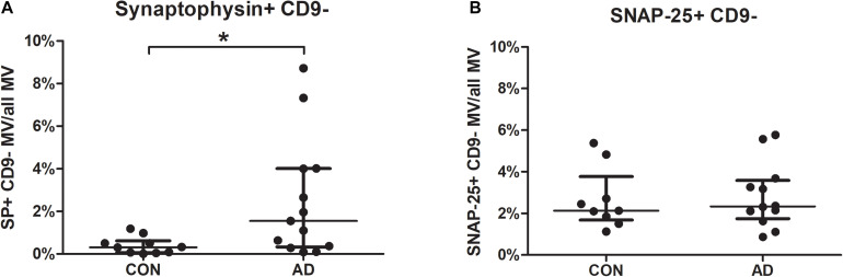
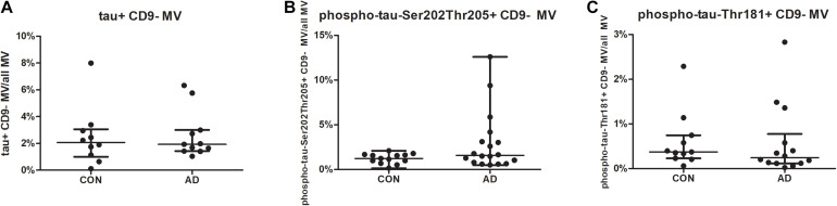
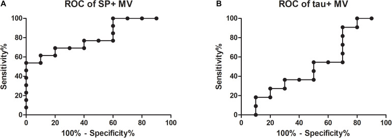
The percentages of synaptophysin-bearing microvesicles were significantly higher in the cerebrospinal fluid of patients with Alzheimer's disease than in the CSF of non-inflammatory neurological disease controls. Tau, phospho-tau-Thr181, phospho-tau-Ser202Thr205, and SNAP-25 did not differ between the groups. The percentages of synaptophysin-bearing vesicles distinguished patients with Alzheimer's disease from the controls (AUC = 0.81).
The loss of synapses in Alzheimer's disease may be reflected by synaptophysin-bearing microvesicles in the cerebrospinal fluid. Future studies are needed to investigate the possibility of using these MVs as a marker to determine the activity of Alzheimer's disease.
在阿尔茨海默病中,症状的严重程度与突触密度的丧失以及病理性过度磷酸化tau蛋白的扩散有关。已确立的脑脊液标志物Aβ、tau蛋白和磷酸化tau蛋白反映了阿尔茨海默病的组织病理学特征,但不能表明疾病进展。此类标志物特别受关注,尤其是对于疾病修饰药物的试验。微泡由应激细胞产生,并反映其起源细胞的部分代谢情况。因此,我们研究了脑脊液中神经元来源的微泡。
我们使用流式细胞术分析了19例阿尔茨海默病患者和15例非炎性神经系统疾病对照者脑脊液中携带tau蛋白、磷酸化tau蛋白-Thr181、磷酸化tau蛋白-Ser202Thr205、突触素和SNAP-25的微泡。
阿尔茨海默病患者脑脊液中携带突触素的微泡百分比显著高于非炎性神经系统疾病对照者的脑脊液。tau蛋白、磷酸化tau蛋白-Thr181、磷酸化tau蛋白-Ser202Thr205和SNAP-25在两组之间无差异。携带突触素的微泡百分比可将阿尔茨海默病患者与对照者区分开来(曲线下面积=0.81)。
阿尔茨海默病中突触的丧失可能由脑脊液中携带突触素的微泡反映出来。未来需要进行研究,以探讨将这些微泡用作确定阿尔茨海默病活性标志物的可能性。