Division of Cardiovascular Medicine, Radcliffe Department of Medicine, University of Oxford, Oxford, UK.
Acute Vascular Imaging Centre, Investigational Medicine, University of Oxford, Oxford, UK.
Br J Pharmacol. 2021 Nov;178(21):4270-4290. doi: 10.1111/bph.15634. Epub 2021 Sep 23.
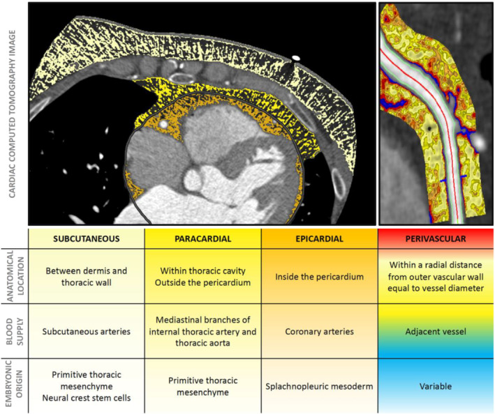
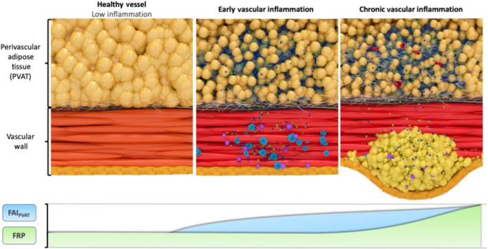
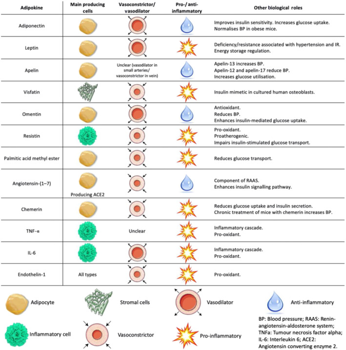
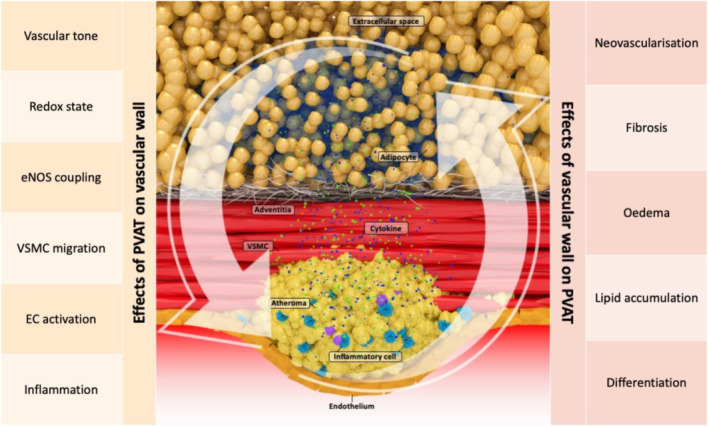
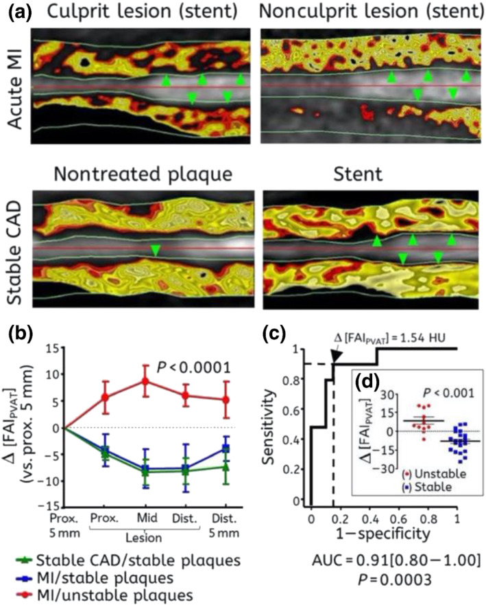
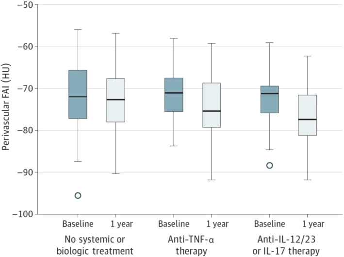
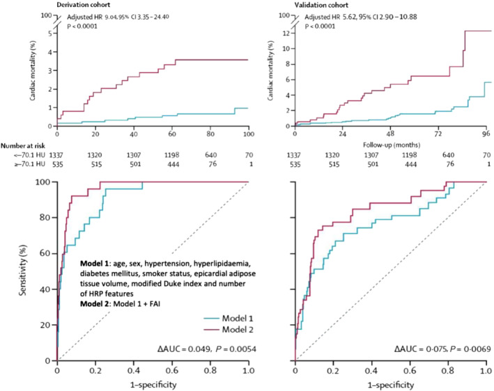
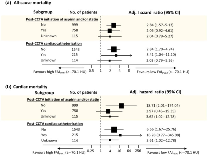
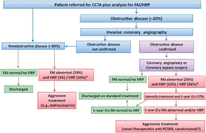
Imaging in medicine has been revolutionised by technological, computational and research advances over the past decades. Computed tomography (CT), in particular, has seen rapid evolution especially in the field of cardiovascular non-invasive imaging. It is being recognised as the first-line tool for the assessment of stable and unstable disease with diagnostic, prognostic and re-stratification potential. Vascular inflammation is a key component of the atherosclerotic process and has been shown to induce molecular, transcriptional and structural changes to perivascular adipose tissue (PVAT). Being a diverse structure itself, PVAT surrounds the human vessels and is characterised by a highly rich secretome, including, amongst others, adipokines, cytokines, gaseous messengers and miRNAs It is implicated in a bidirectional interplay with the adjacent vascular wall, affecting and being affected by aspects of its biology, mainly inflammation. In this review, we discuss the current status of cardiac CT in imaging vascular inflammation through PVAT phenotyping. LINKED ARTICLES: This article is part of a themed issue on Molecular imaging - visual themed issue. To view the other articles in this section visit http://onlinelibrary.wiley.com/doi/10.1111/bph.v178.21/issuetoc.
在过去几十年中,医学影像学在技术、计算和研究方面取得了革命性的进展。特别是计算机断层扫描(CT),特别是在心血管无创成像领域,已经得到了快速发展。它被认为是评估稳定和不稳定疾病的一线工具,具有诊断、预后和再分层的潜力。血管炎症是动脉粥样硬化过程的一个关键组成部分,已被证明会诱导血管周围脂肪组织(PVAT)的分子、转录和结构变化。作为一个多样化的结构,PVAT 环绕着人体血管,其特征是富含丰富的分泌组,包括脂肪因子、细胞因子、气体信使和 miRNAs。它与相邻的血管壁之间存在双向相互作用,影响和被其生物学的各个方面所影响,主要是炎症。在这篇综述中,我们讨论了通过 PVAT 表型成像评估血管炎症的心脏 CT 的现状。相关文章:本文是分子成像-视觉主题专辑的一部分。要查看本节中的其他文章,请访问 http://onlinelibrary.wiley.com/doi/10.1111/bph.v178.21/issuetoc.