Department of Pathology and Laboratory Medicine, Robert Wood Johnson Medical School, Rutgers, The State University of New Jersey, Piscataway, NJ 08854, USA.
OptoVibronex, LLC., Allentown, PA 18104, USA.
Biomolecules. 2021 Jul 12;11(7):1018. doi: 10.3390/biom11071018.
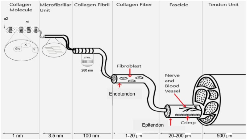
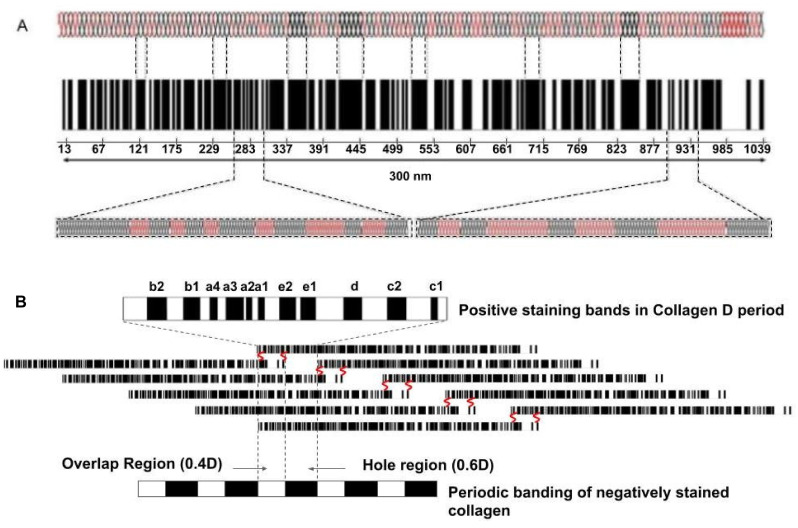
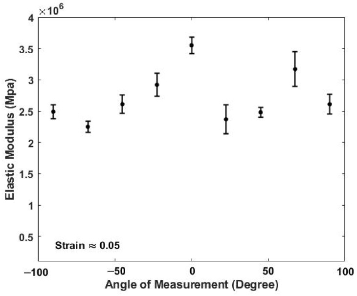
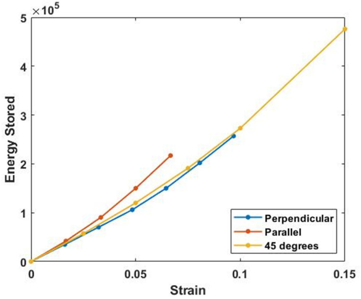
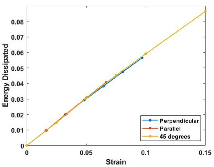
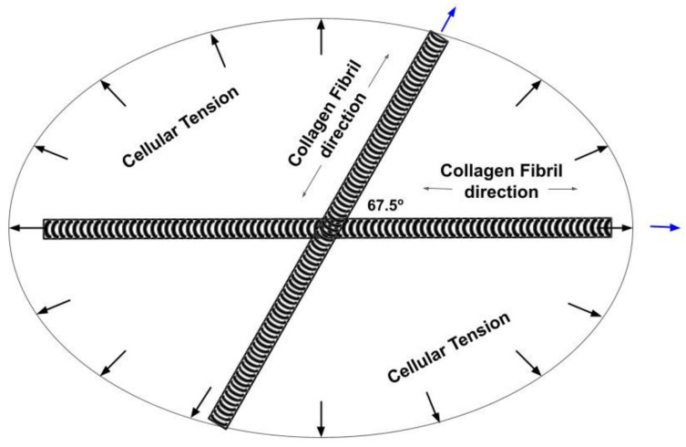
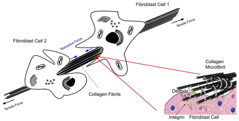
Collagen and proteoglycans work in unison in the ECM to bear loads, store elastic energy and then dissipate excess energy to avoid tissue fatigue and premature mechanical failure. While collagen fibers store elastic energy by stretching the flexible regions in the triple helix, they do so by lowering their free energy through a reduction in the entropy and a decrease in charge-charge repulsion. Entropic increases occur when the load is released that drive the reversibility of the process and transmission of excess energy. Energy is dissipated by sliding of collagen fibrils by each other with the aid of decorin molecules that reside on the d and e bands of the native D repeat pattern. Fluid flow from the hydration layer associated with the decorin and collagen fibrils hydraulically dissipates energy during sliding. The deformation is reversed by osmotic forces that cause fluid to reform a hydration shell around the collagen fibrils when the loads are removed. In this paper a model is presented describing the organization of collagen fibers in the skin and cell-collagen mechanical relationships that exist based on non-invasive measurements made using vibrational optical coherence tomography. It is proposed that under external stress, collagen fibers form a tensional network in the plane of the skin. Collagen fiber tension along with forces generated by fibroblasts exerted on collagen fibers lead to an elastic modulus that is almost uniform throughout the plane of the skin. Tensile forces acting on cells and tissues may provide a baseline for stimulation of normal mechanotransduction. We hypothesize that during aging, changes in cellular metabolism, cell-collagen interactions and light and UV light exposure cause down regulation of mechanotransduction and tissue metabolism leading to tissue atrophy.
胶原蛋白和蛋白聚糖在细胞外基质中协同工作,以承受负荷、储存弹性能量,然后耗散多余的能量,以避免组织疲劳和过早的机械失效。虽然胶原纤维通过拉伸三螺旋中的柔性区域来储存弹性能量,但它们通过降低熵和减少电荷-电荷排斥来降低自由能。当负载释放时,熵的增加会驱动过程的可逆性和多余能量的传递。能量通过借助存在于天然 D 重复模式的 d 和 e 带中的饰胶蛋白分子的胶原原纤维的滑动而耗散。与饰胶蛋白和胶原原纤维相关的水合层中的流体流动在滑动过程中耗散能量。当负载移除时,渗透压会使流体重新形成胶原原纤维周围的水合壳,从而使变形反转。本文提出了一个模型,描述了皮肤中胶原纤维的组织以及基于使用振动光学相干断层扫描进行的非侵入性测量所存在的细胞-胶原机械关系。据提议,在外部应力下,胶原纤维在皮肤的平面内形成张紧网络。胶原纤维张力以及成纤维细胞施加在胶原纤维上的力导致弹性模量在皮肤平面内几乎均匀。作用于细胞和组织的拉伸力可能为正常机械转导的刺激提供基线。我们假设,在衰老过程中,细胞代谢、细胞-胶原相互作用以及光和紫外线暴露的变化会导致机械转导和组织代谢的下调,从而导致组织萎缩。