Brown Wendy E, Huang Brian J, Hu Jerry C, Athanasiou Kyriacos A
Department of Biomedical Engineering, University of California Irvine, 3120 Natural Sciences II, Irvine, CA, USA.
Integrative Stem Cell Center, China Medical University Hospital, Taichung, Taiwan.
NPJ Regen Med. 2021 Aug 6;6(1):42. doi: 10.1038/s41536-021-00152-0.
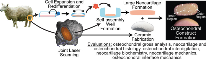
Despite the prevalence of large (>5 cm) articular cartilage defects involving underlying bone, current tissue-engineered therapies only address small defects. Tissue-engineered, anatomically shaped, native-like implants may address the need for off-the-shelf, tissue-repairing therapies for large cartilage lesions. This study fabricated an osteochondral construct of translationally relevant geometry with robust functional properties. Scaffold-free, self-assembled neocartilage served as the chondral phase, and porous hydroxyapatite served as the osseous phase of the osteochondral constructs. Constructs in the shape and size of an ovine femoral condyle (31 × 14 mm) were assembled at day 4 (early) or day 10 (late) of neocartilage maturation. Early osteochondral assembly increased the interfacial interdigitation depth by 244%, interdigitation frequency by 438%, interfacial shear modulus by 243-fold, and ultimate interfacial shear strength by 4.9-fold, compared to late assembly. Toward the development of a bioprosthesis for the repair of cartilage lesions encompassing up to an entire condylar surface, this study generated a large, anatomically shaped osteochondral construct with robust interfacial mechanical properties and native-like neocartilage interdigitation.
尽管涉及深层骨的大尺寸(>5厘米)关节软骨缺损普遍存在,但目前的组织工程疗法仅针对小尺寸缺损。组织工程化的、符合解剖形状的、类似天然组织的植入物可能满足对大尺寸软骨损伤的现货供应型组织修复疗法的需求。本研究制造了一种具有强大功能特性的、与转化相关几何形状的骨软骨构建体。无支架自组装新软骨作为软骨相,多孔羟基磷灰石作为骨软骨构建体的骨相。在新软骨成熟的第4天(早期)或第10天(晚期)组装成羊股骨髁形状和尺寸(31×14毫米)的构建体。与晚期组装相比,早期骨软骨组装使界面相互交错深度增加了244%,相互交错频率增加了438%,界面剪切模量增加了243倍,极限界面剪切强度增加了4.9倍。为了开发用于修复多达整个髁表面的软骨损伤的生物假体,本研究生成了一种具有强大界面力学性能和类似天然新软骨相互交错的大尺寸、符合解剖形状的骨软骨构建体。