Department of Biomedical Engineering, Washington University in St. Louis, St. Louis, Missouri.
Department of Radiology, Mallinckrodt Institute of Radiology, Washington University School of Medicine, St. Louis, Missouri.
J Nucl Med. 2022 Apr;63(4):591-597. doi: 10.2967/jnumed.121.262270. Epub 2021 Aug 12.
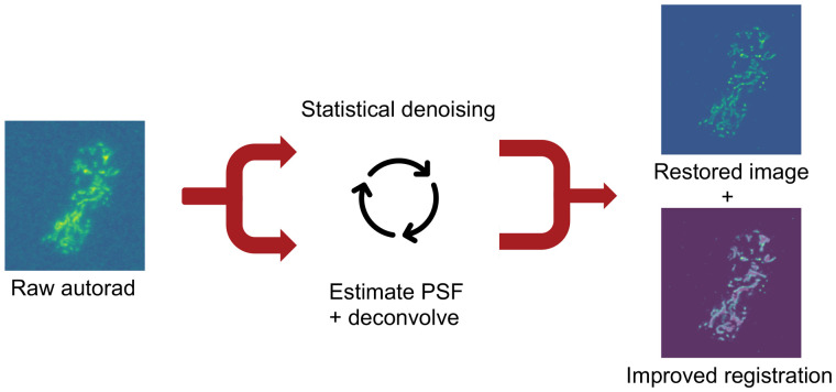
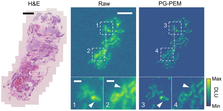
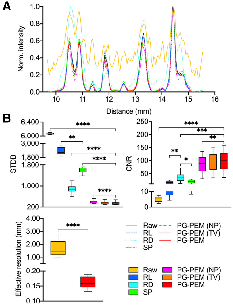
Digital autoradiography (DAR) is a powerful tool to quantitatively determine the distribution of a radiopharmaceutical within a tissue section and is widely used in drug discovery and development. However, the low image resolution and significant background noise can result in poor correlation, even errors, between radiotracer distribution, anatomic structure, and molecular expression profiles. Differing from conventional optical systems, the point-spread function in DAR is determined by properties of radioisotope decay, phosphor, and digitizer. Calibration of an experimental point-spread function a priori is difficult, prone to error, and impractical. We have developed a content-adaptive restoration algorithm to address these problems. We model the DAR imaging process using a mixed Poisson-gaussian model and blindly restore the image by a penalized maximum-likelihood expectation-maximization algorithm (PG-PEM). PG-PEM implements a patch-based estimation algorithm with density-based spatial clustering of applications with noise to estimate noise parameters and uses L2 and Hessian Frebonius norms as regularization functions to improve performance. First, PG-PEM outperformed other restoration algorithms at the denoising task ( 0.01). Next, we implemented PG-PEM on preclinical DAR images (F-FDG, treated mouse tumor and heart; F-NaF, treated mouse femur) and clinical DAR images (bone biopsy sections from RaCl-treated castration-resistant prostate cancer patients). DAR images restored by PG-PEM of all samples achieved a significantly higher effective resolution and contrast-to-noise ratio and a lower SD of background ( 0.0001). Additionally, by comparing the registration results between the clinical DAR images and the segmented bone masks from the corresponding histologic images, we found that the radiopharmaceutical distribution was significantly improved ( 0.0001). PG-PEM is able to increase resolution and contrast while robustly accounting for DAR noise and demonstrates the capacity to be widely implemented to improve preclinical and clinical DAR imaging of radiopharmaceutical distribution.
数字放射自显影术(DAR)是一种强大的工具,可定量确定放射性药物在组织切片中的分布,广泛用于药物发现和开发。然而,低图像分辨率和显著的背景噪声可能导致放射性示踪剂分布、解剖结构和分子表达谱之间相关性差,甚至出现错误。与传统的光学系统不同,DAR 中的点扩散函数由放射性同位素衰变、荧光体和数字化仪的特性决定。实验性点扩散函数的预先校准是困难的、容易出错的且不切实际的。我们开发了一种内容自适应恢复算法来解决这些问题。我们使用混合泊松-高斯模型对 DAR 成像过程进行建模,并通过惩罚最大似然期望最大化算法(PG-PEM)盲目恢复图像。PG-PEM 实现了基于补丁的估计算法,该算法具有基于密度的空间聚类应用噪声以估计噪声参数,并使用 L2 和 Hessian Frebonius 范数作为正则化函数来提高性能。首先,PG-PEM 在去噪任务上优于其他恢复算法(P<0.01)。接下来,我们在临床前 DAR 图像(F-FDG,治疗的小鼠肿瘤和心脏;F-NaF,治疗的小鼠股骨)和临床 DAR 图像(接受 RaCl 治疗的去势抵抗性前列腺癌患者的骨活检切片)上实现了 PG-PEM。所有样本的 PG-PEM 恢复的 DAR 图像均实现了更高的有效分辨率、更高的对比度噪声比和更低的背景标准差(P<0.0001)。此外,通过比较临床 DAR 图像和相应组织学图像的分割骨掩模之间的配准结果,我们发现放射性药物分布得到了显著改善(P<0.0001)。PG-PEM 能够在稳健地考虑 DAR 噪声的同时提高分辨率和对比度,并展示了在改善放射性药物分布的临床前和临床 DAR 成像方面广泛实施的能力。