Laboratory of Clinical Nuclear Medicine, Department of Nuclear Medicine, West China Hospital, Sichuan University, No. 37 Guo Xue Alley, Chengdu, 610041, China.
Machine Intelligence Laboratory, College of Computer Science, Sichuan University, Chengdu, 610065, China.
BMC Med Imaging. 2021 Sep 4;21(1):131. doi: 10.1186/s12880-021-00662-9.
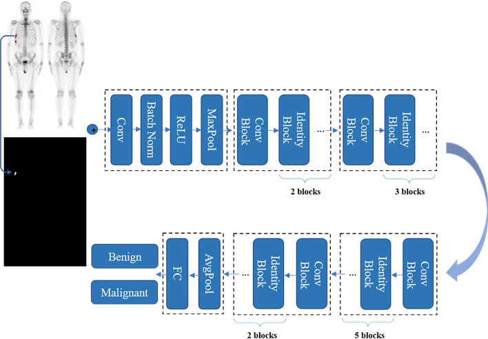
We aimed to construct an artificial intelligence (AI) guided identification of suspicious bone metastatic lesions from the whole-body bone scintigraphy (WBS) images by convolutional neural networks (CNNs).
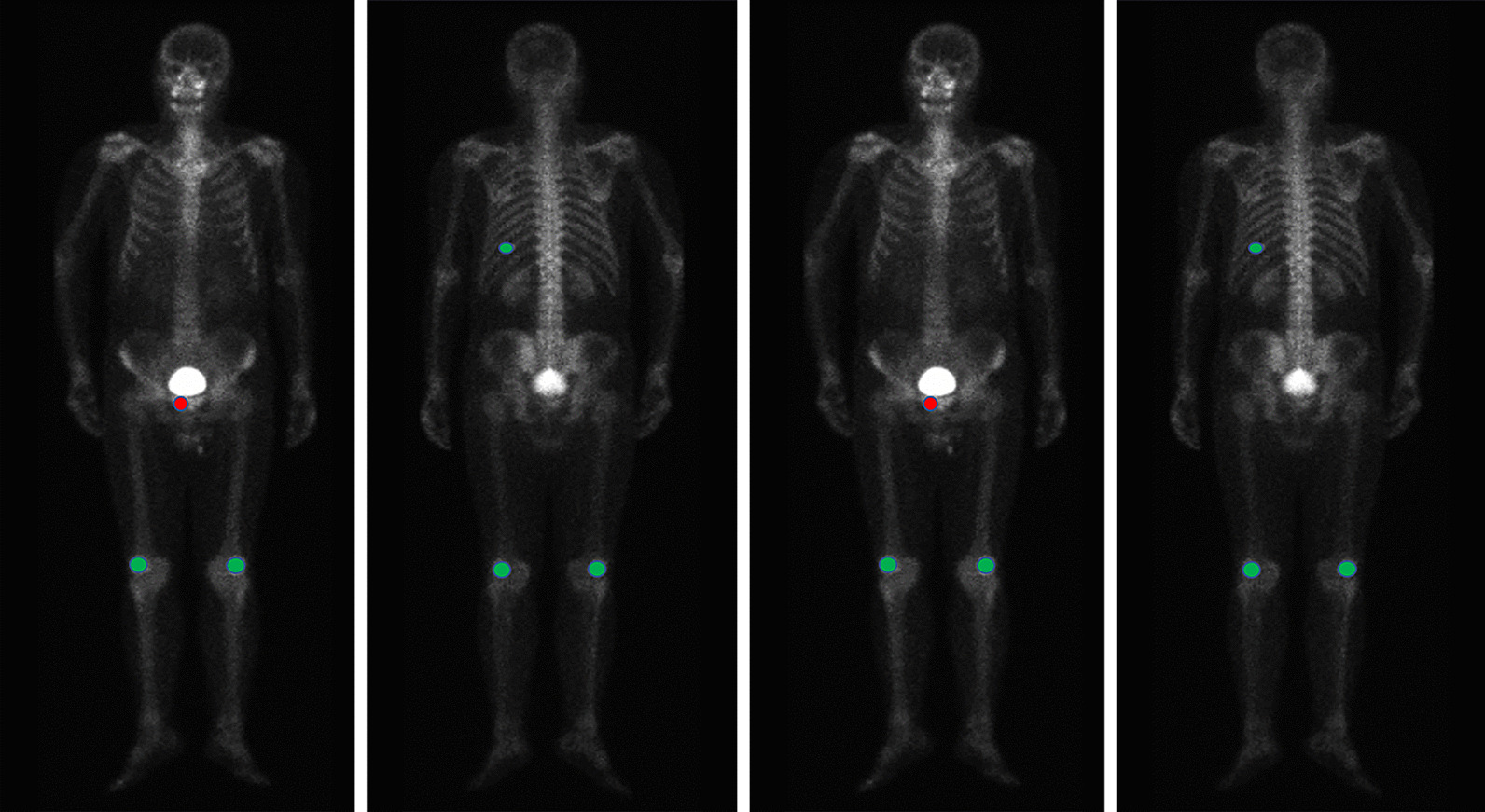
We retrospectively collected the Tc-MDP WBS images with confirmed bone lesions from 3352 patients with malignancy. 14,972 bone lesions were delineated manually by physicians and annotated as benign and malignant. The lesion-based differentiating performance of the proposed network was evaluated by fivefold cross validation, and compared with the other three popular CNN architectures for medical imaging. The average sensitivity, specificity, accuracy and the area under receiver operating characteristic curve (AUC) were calculated. To delve the outcomes of this study, we conducted subgroup analyses, including lesion burden number and tumor type for the classifying ability of the CNN.
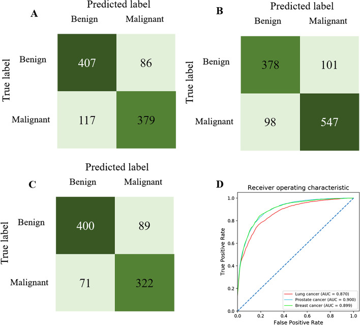
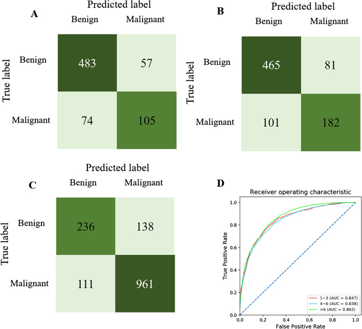
In the fivefold cross validation, our proposed network reached the best average accuracy (81.23%) in identifying suspicious bone lesions compared with InceptionV3 (80.61%), VGG16 (81.13%) and DenseNet169 (76.71%). Additionally, the CNN model's lesion-based average sensitivity and specificity were 81.30% and 81.14%, respectively. Based on the lesion burden numbers of each image, the area under the receiver operating characteristic curve (AUC) was 0.847 in the few group (lesion number n ≤ 3), 0.838 in the medium group (n = 4-6), and 0.862 in the extensive group (n > 6). For the three major primary tumor types, the CNN-based lesion identifying AUC value was 0.870 for lung cancer, 0.900 for prostate cancer, and 0.899 for breast cancer.
The CNN model suggests potential in identifying suspicious benign and malignant bone lesions from whole-body bone scintigraphic images.
我们旨在通过卷积神经网络(CNN)构建一种从全身骨闪烁显像(WBS)图像中识别可疑骨转移病变的人工智能(AI)引导方法。
我们回顾性收集了 3352 例恶性肿瘤患者经 Tc-MDP WBS 图像证实的骨病变。由医师手动描绘 14972 个骨病变,并将其标注为良性和恶性。通过五重交叉验证评估所提出的网络的基于病变的区分性能,并与其他三种用于医学成像的流行 CNN 架构进行比较。计算平均灵敏度、特异性、准确性和受试者工作特征曲线(ROC)下的面积(AUC)。为深入研究本研究的结果,我们进行了亚组分析,包括病变负担数量和肿瘤类型,以评估 CNN 的分类能力。
在五重交叉验证中,与 InceptionV3(80.61%)、VGG16(81.13%)和 DenseNet169(76.71%)相比,我们提出的网络在识别可疑骨病变方面达到了最佳的平均准确性(81.23%)。此外,CNN 模型的基于病变的平均灵敏度和特异性分别为 81.30%和 81.14%。基于每个图像的病变负担数量,ROC 曲线下的面积(AUC)在病变数量较少的组(病变数量 n≤3)中为 0.847,在病变数量中等的组(n=4-6)中为 0.838,在病变数量较多的组(n>6)中为 0.862。对于三种主要的原发性肿瘤类型,基于 CNN 的病变识别 AUC 值分别为肺癌 0.870、前列腺癌 0.900 和乳腺癌 0.899。
CNN 模型有望从全身骨闪烁显像图像中识别可疑的良性和恶性骨病变。