School of Mathematics and Computer Science, Northwest Minzu University, Lanzhou, Gansu, China.
Key Laboratory of China's Ethnic Languages and Information Technology of Ministry of Education, Northwest Minzu University, Lanzhou, Gansu, China.
PLoS One. 2020 Dec 3;15(12):e0243253. doi: 10.1371/journal.pone.0243253. eCollection 2020.
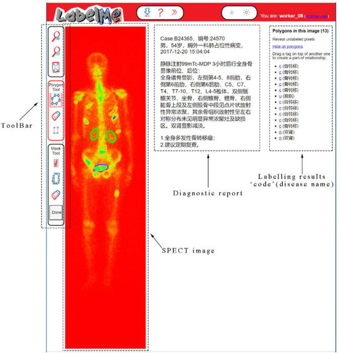
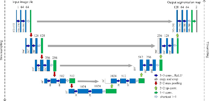
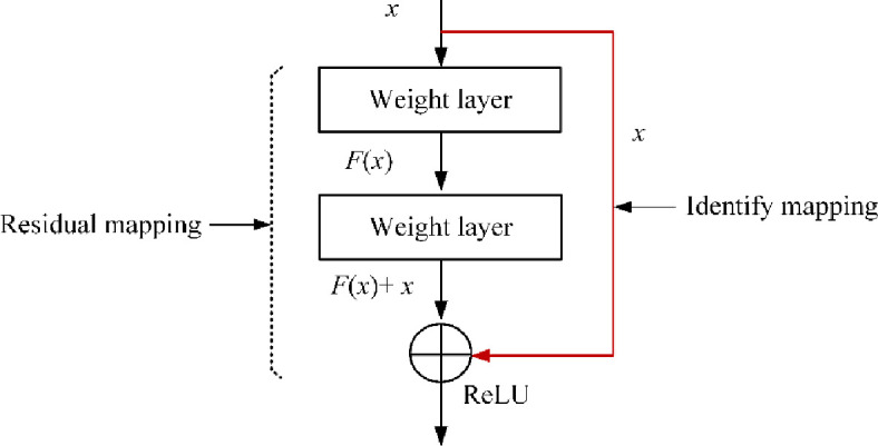
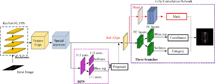
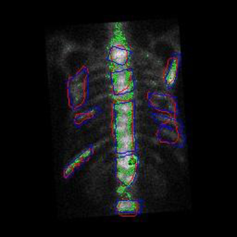
SPECT imaging has been identified as an effective medical modality for diagnosis, treatment, evaluation and prevention of a range of serious diseases and medical conditions. Bone SPECT scan has the potential to provide more accurate assessment of disease stage and severity. Segmenting hotspot in bone SPECT images plays a crucial role to calculate metrics like tumor uptake and metabolic tumor burden. Deep learning techniques especially the convolutional neural networks have been widely exploited for reliable segmentation of hotspots or lesions, organs and tissues in the traditional structural medical images (i.e., CT and MRI) due to their ability of automatically learning the features from images in an optimal way. In order to segment hotspots in bone SPECT images for automatic assessment of metastasis, in this work, we develop several deep learning based segmentation models. Specifically, each original whole-body bone SPECT image is processed to extract the thorax area, followed by image mirror, translation and rotation operations, which augments the original dataset. We then build segmentation models based on two commonly-used famous deep networks including U-Net and Mask R-CNN by fine-tuning their structures. Experimental evaluation conducted on a group of real-world bone SEPCT images reveals that the built segmentation models are workable on identifying and segmenting hotspots of metastasis in bone SEPCT images, achieving a value of 0.9920, 0.7721, 0.6788 and 0.6103 for PA (accuracy), CPA (precision), Rec (recall) and IoU, respectively. Finally, we conclude that the deep learning technology have the huge potential to identify and segment hotspots in bone SPECT images.
SPECT 成像已被确定为诊断、治疗、评估和预防一系列严重疾病和医疗状况的有效医学手段。骨 SPECT 扫描有可能提供更准确的疾病分期和严重程度评估。在骨 SPECT 图像中分割热点对于计算肿瘤摄取和代谢肿瘤负担等指标至关重要。深度学习技术,特别是卷积神经网络,由于其能够以最佳方式自动从图像中学习特征,因此已被广泛用于传统结构医学图像(即 CT 和 MRI)中热点或病变、器官和组织的可靠分割。为了对转移进行自动评估,在这项工作中,我们开发了几种基于深度学习的分割模型。具体来说,每个原始全身骨 SPECT 图像都经过处理以提取胸部区域,然后进行图像镜像、平移和旋转操作,从而扩充原始数据集。然后,我们基于 U-Net 和 Mask R-CNN 这两个常用的著名深度学习网络构建分割模型,通过微调它们的结构来进行训练。在一组真实的骨 SPECT 图像上进行的实验评估表明,所构建的分割模型可用于识别和分割骨 SPECT 图像中的转移热点,其 PA(准确率)、CPA(精确率)、Rec(召回率)和 IoU 的值分别为 0.9920、0.7721、0.6788 和 0.6103。最后,我们得出结论,深度学习技术具有识别和分割骨 SPECT 图像中热点的巨大潜力。