• 文献检索
  • 文档翻译
  • 深度研究
  • 学术资讯
  • Suppr Zotero 插件Zotero 插件
  • 邀请有礼
  • 套餐&价格
  • 历史记录
应用&插件
Suppr Zotero 插件Zotero 插件浏览器插件Mac 客户端Windows 客户端微信小程序
定价
高级版会员购买积分包购买API积分包
服务
文献检索文档翻译深度研究API 文档MCP 服务
关于我们
关于 Suppr公司介绍联系我们用户协议隐私条款
关注我们

Suppr 超能文献

核心技术专利:CN118964589B侵权必究
粤ICP备2023148730 号-1Suppr @ 2026

文献检索

告别复杂PubMed语法,用中文像聊天一样搜索,搜遍4000万医学文献。AI智能推荐,让科研检索更轻松。

立即免费搜索

文件翻译

保留排版,准确专业,支持PDF/Word/PPT等文件格式,支持 12+语言互译。

免费翻译文档

深度研究

AI帮你快速写综述,25分钟生成高质量综述,智能提取关键信息,辅助科研写作。

立即免费体验

影像检查在恶性胸膜间皮瘤诊断、分期及治疗反应评估中的新进展

New Updates of the Imaging Role in Diagnosis, Staging, and Response Treatment of Malignant Pleural Mesothelioma.

作者信息

Romei Chiara, Fanni Salvatore Claudio, Volpi Federica, Milazzo Alessio, D'Amore Caterina Aida, Colligiani Leonardo, Neri Emanuele, De Liperi Annalisa, Stella Giulia Maria, Bortolotto Chandra

机构信息

2nd Radiology Unit, Radiology Department, Pisa University Hospital, 56124 Pisa, Italy.

Department of Translational Research, Academic Radiology, University of Pisa, 56124 Pisa, Italy.

出版信息

Cancers (Basel). 2021 Aug 30;13(17):4377. doi: 10.3390/cancers13174377.

DOI:10.3390/cancers13174377
PMID:34503186
原文链接:https://pmc.ncbi.nlm.nih.gov/articles/PMC8430786/
Abstract

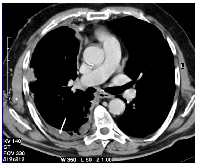
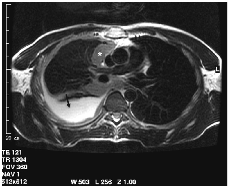
Malignant pleural mesothelioma is a rare neoplasm with poor prognosis. CT is the first imaging technique used for diagnosis, staging, and assessment of therapy response. Although, CT has intrinsic limitations due to low soft tissue contrast and the current staging system as well as criteria for evaluating response, it does not consider the complex growth pattern of this tumor. Computer-based methods have proven their potentiality in diagnosis, staging, prognosis, and assessment of therapy response; moreover, computer-based methods can make feasible tasks like segmentation that would otherwise be impracticable. MRI, thanks to its high soft tissue contrast evaluation of contrast enhancement and through diffusion-weighted-images, could replace CT in many clinical settings.

摘要

恶性胸膜间皮瘤是一种预后较差的罕见肿瘤。CT是用于诊断、分期及评估治疗反应的首选成像技术。尽管CT由于软组织对比度低以及当前的分期系统和评估反应的标准而存在固有局限性,但它并未考虑该肿瘤复杂的生长模式。基于计算机的方法已在诊断、分期、预后及治疗反应评估中证明了其潜力;此外,基于计算机的方法可完成如分割等原本不可行的任务。MRI凭借其对对比增强的高软组织对比度评估以及通过扩散加权成像,在许多临床情况下可替代CT。

https://cdn.ncbi.nlm.nih.gov/pmc/blobs/1414/8430786/225ddfd13db8/cancers-13-04377-g003.jpg
https://cdn.ncbi.nlm.nih.gov/pmc/blobs/1414/8430786/f13572d724ea/cancers-13-04377-g001.jpg
https://cdn.ncbi.nlm.nih.gov/pmc/blobs/1414/8430786/8edb222fd67d/cancers-13-04377-g002.jpg
https://cdn.ncbi.nlm.nih.gov/pmc/blobs/1414/8430786/225ddfd13db8/cancers-13-04377-g003.jpg
https://cdn.ncbi.nlm.nih.gov/pmc/blobs/1414/8430786/f13572d724ea/cancers-13-04377-g001.jpg
https://cdn.ncbi.nlm.nih.gov/pmc/blobs/1414/8430786/8edb222fd67d/cancers-13-04377-g002.jpg
https://cdn.ncbi.nlm.nih.gov/pmc/blobs/1414/8430786/225ddfd13db8/cancers-13-04377-g003.jpg

相似文献

1
New Updates of the Imaging Role in Diagnosis, Staging, and Response Treatment of Malignant Pleural Mesothelioma.影像检查在恶性胸膜间皮瘤诊断、分期及治疗反应评估中的新进展
Cancers (Basel). 2021 Aug 30;13(17):4377. doi: 10.3390/cancers13174377.
2
Imaging in pleural mesothelioma: a review of the 11th International Conference of the International Mesothelioma Interest Group.胸膜间皮瘤的影像学:第 11 届国际间皮瘤兴趣小组国际会议综述。
Lung Cancer. 2013 Nov;82(2):190-6. doi: 10.1016/j.lungcan.2013.08.005. Epub 2013 Aug 15.
3
Morphologic and functional imaging of malignant pleural mesothelioma.恶性胸膜间皮瘤的形态学和功能成像
Eur J Radiol. 2007 Dec;64(3):356-66. doi: 10.1016/j.ejrad.2007.08.010. Epub 2007 Oct 22.
4
Staging and response to therapy of malignant pleural mesothelioma.恶性胸膜间皮瘤的分期及对治疗的反应
Lung Cancer. 2004 Aug;45 Suppl 1:S59-61. doi: 10.1016/j.lungcan.2004.04.013.
5
Whole-Body MRI: Comparison of Its Capability for TNM Staging of Malignant Pleural Mesothelioma With That of Coregistered PET/MRI, Integrated FDG PET/CT, and Conventional Imaging.全身 MRI 与核素融合 PET/MRI、一体化 FDG PET/CT 和常规成像在恶性胸膜间皮瘤 TNM 分期中的比较。
AJR Am J Roentgenol. 2019 Feb;212(2):311-319. doi: 10.2214/AJR.18.20111. Epub 2018 Dec 4.
6
The role of new imaging techniques in diagnosis and staging of malignant pleural mesothelioma.新成像技术在恶性胸膜间皮瘤诊断及分期中的作用
Curr Opin Oncol. 2003 Mar;15(2):131-8. doi: 10.1097/00001622-200303000-00003.
7
Diagnostic accuracy of sequential co-registered PET+MR in comparison to PET/CT in local thoracic staging of malignant pleural mesothelioma.序贯同机融合PET+MR与PET/CT在恶性胸膜间皮瘤局部胸部分期中的诊断准确性比较
Lung Cancer. 2016 Apr;94:40-5. doi: 10.1016/j.lungcan.2016.01.017. Epub 2016 Jan 28.
8
Optimal gross tumor volume definition in lung-sparing intensity modulated radiotherapy for pleural mesothelioma: an in silico study.胸膜间皮瘤肺保留调强放射治疗中最佳肿瘤总体积定义:一项计算机模拟研究
Acta Oncol. 2016 Dec;55(12):1450-1455. doi: 10.1080/0284186X.2016.1234066. Epub 2016 Oct 12.
9
[MRT, CT and sonography in the preoperative assessment of the primary tumor spread in malignant pleural mesothelioma].[磁共振成像、计算机断层扫描及超声检查在恶性胸膜间皮瘤原发肿瘤播散术前评估中的应用]
Rofo. 1999 Apr;170(4):365-70. doi: 10.1055/s-2007-1011055.
10
Use of imaging in the management of malignant pleural mesothelioma.影像学在恶性胸膜间皮瘤管理中的应用。
Clin Radiol. 2005 Dec;60(12):1237-47. doi: 10.1016/j.crad.2005.05.015.

引用本文的文献

1
A Diagnostic Approach to Malignant Pleural Mesothelioma.恶性胸膜间皮瘤的诊断方法
Pulm Ther. 2025 Jun 11. doi: 10.1007/s41030-025-00301-6.
2
Advanced imaging techniques and artificial intelligence in pleural diseases: a narrative review.胸膜疾病中的先进成像技术与人工智能:一篇叙述性综述
Eur Respir Rev. 2025 Apr 2;34(176). doi: 10.1183/16000617.0263-2024. Print 2025 Apr.
3
Unusual Cause of Diffuse Pleural Thickening (Malignant Pleural Mesothelioma Mimic): Undifferentiated Epithelioid Malignant Neoplasm.弥漫性胸膜增厚的罕见病因(类恶性胸膜间皮瘤):未分化上皮样恶性肿瘤

本文引用的文献

1
A comparison between MRI and CT in the assessment of primary tumour volume in mesothelioma.磁共振成像(MRI)与计算机断层扫描(CT)在评估间皮瘤原发肿瘤体积方面的比较。
Lung Cancer. 2020 Dec;150:12-20. doi: 10.1016/j.lungcan.2020.09.025. Epub 2020 Oct 1.
2
FDG PET versus CT radiomics to predict outcome in malignant pleural mesothelioma patients.18F-氟脱氧葡萄糖正电子发射断层扫描(FDG PET)与计算机断层扫描(CT)影像组学在预测恶性胸膜间皮瘤患者预后中的应用比较
EJNMMI Res. 2020 Jul 13;10(1):81. doi: 10.1186/s13550-020-00669-3.
3
ERS/ESTS/EACTS/ESTRO guidelines for the management of malignant pleural mesothelioma.
Oman Med J. 2024 Jul 31;39(4):e657. doi: 10.5001/omj.2024.27. eCollection 2024 Jul.
4
Precision matters: the value of PET/CT and PET/MRI in the clinical management of cervical cancer.精准至关重要:PET/CT与PET/MRI在宫颈癌临床管理中的价值
Strahlenther Onkol. 2025 May;201(5):507-518. doi: 10.1007/s00066-024-02294-8. Epub 2024 Sep 27.
5
Malignant Pleural Mesothelioma: Staging and Radiological Response Criteria in Patients Treated with Immune Checkpoint Inhibitors.恶性胸膜间皮瘤:免疫检查点抑制剂治疗患者的分期和影像学反应标准。
Target Oncol. 2024 Jan;19(1):13-28. doi: 10.1007/s11523-023-01017-w. Epub 2023 Dec 8.
6
A first look into radiomics application in testicular imaging: A systematic review.睾丸成像中放射组学应用的初步研究:一项系统综述。
Front Radiol. 2023 Apr 17;3:1141499. doi: 10.3389/fradi.2023.1141499. eCollection 2023.
7
Pleural Neoplasms-What Could MRI Change?胸膜肿瘤——MRI能带来哪些改变?
Cancers (Basel). 2023 Jun 20;15(12):3261. doi: 10.3390/cancers15123261.
8
Role of artificial intelligence in oncologic emergencies: a narrative review.人工智能在肿瘤急症中的作用:一项叙述性综述
Explor Target Antitumor Ther. 2023;4(2):344-354. doi: 10.37349/etat.2023.00138. Epub 2023 Apr 28.
9
Imaging Diagnosis of Hepatocellular Carcinoma: A State-of-the-Art Review.肝细胞癌的影像学诊断:最新进展综述
Diagnostics (Basel). 2023 Feb 8;13(4):625. doi: 10.3390/diagnostics13040625.
10
Managing Malignant Pleural Mesothelioma in the Age of Personalized Medicine: Where Are We and What Is Still Missing?个性化医疗时代的恶性胸膜间皮瘤管理:我们目前的状况及仍存在的不足?
Cancers (Basel). 2022 Nov 11;14(22):5540. doi: 10.3390/cancers14225540.
ERS/ESTS/EACTS/ESTRO 指南:恶性胸膜间皮瘤的管理。
Eur Respir J. 2020 Jun 11;55(6). doi: 10.1183/13993003.00953-2019. Print 2020 Jun.
4
Epidemiology of mesothelioma in the 21 century in Europe and the United States, 40 years after restricted/banned asbestos use.在欧洲和美国限制/禁止使用石棉40年后,21世纪间皮瘤的流行病学情况
Transl Lung Cancer Res. 2020 Feb;9(Suppl 1):S28-S38. doi: 10.21037/tlcr.2019.11.11.
5
Preoperative prediction of unresectability in malignant pleural mesothelioma.恶性胸膜间皮瘤不可切除性的术前预测。
J Thorac Cardiovasc Surg. 2020 Jun;159(6):2512-2520.e1. doi: 10.1016/j.jtcvs.2019.11.035. Epub 2019 Nov 27.
6
Deep learning-based segmentation of malignant pleural mesothelioma tumor on computed tomography scans: application to scans demonstrating pleural effusion.基于深度学习的计算机断层扫描中恶性胸膜间皮瘤肿瘤分割:应用于显示胸腔积液的扫描图像
J Med Imaging (Bellingham). 2020 Jan;7(1):012705. doi: 10.1117/1.JMI.7.1.012705. Epub 2020 Jan 29.
7
Mesothelioma: Scientific clues for prevention, diagnosis, and therapy.间皮瘤:预防、诊断和治疗的科学线索。
CA Cancer J Clin. 2019 Sep;69(5):402-429. doi: 10.3322/caac.21572. Epub 2019 Jul 8.
8
Diffusion-Weighted Imaging Can Differentiate between Malignant and Benign Pleural Diseases.扩散加权成像可鉴别恶性和良性胸膜疾病。
Cancers (Basel). 2019 Jun 12;11(6):811. doi: 10.3390/cancers11060811.
9
Deep convolutional neural networks for the automated segmentation of malignant pleural mesothelioma on computed tomography scans.用于在计算机断层扫描上自动分割恶性胸膜间皮瘤的深度卷积神经网络。
J Med Imaging (Bellingham). 2018 Jul;5(3):034503. doi: 10.1117/1.JMI.5.3.034503. Epub 2018 Sep 24.
10
Dynamic contrast-enhanced MRI of malignant pleural mesothelioma: a comparative study of pharmacokinetic models and correlation with mRECIST criteria.动态对比增强 MRI 对恶性胸膜间皮瘤的研究:药代动力学模型的比较研究及其与 mRECIST 标准的相关性。
Cancer Imaging. 2019 Feb 27;19(1):10. doi: 10.1186/s40644-019-0189-5.