Brown Nicholas M, McDonald James F, Sershon Robert A, Hopper Robert H
Department of Orthopaedic Surgery, Loyola University Medical Center, Maywood, IL, USA.
Department of Orthopaedic Surgery, Anderson Orthopaedic Research Institute, Alexandria, VA, USA.
Hip Pelvis. 2021 Sep;33(3):128-139. doi: 10.5371/hp.2021.33.3.128. Epub 2021 Sep 6.
Accurate component placement and restoration of patient anatomy are critical in total hip arthroplasty (THA) surgery. Although intraoperative radiographs are sometimes utilized, it is unclear whether this practice can improve accuracy.
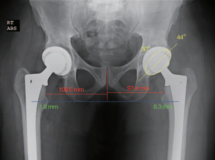
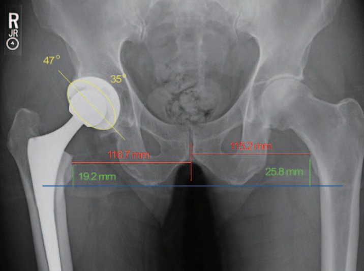
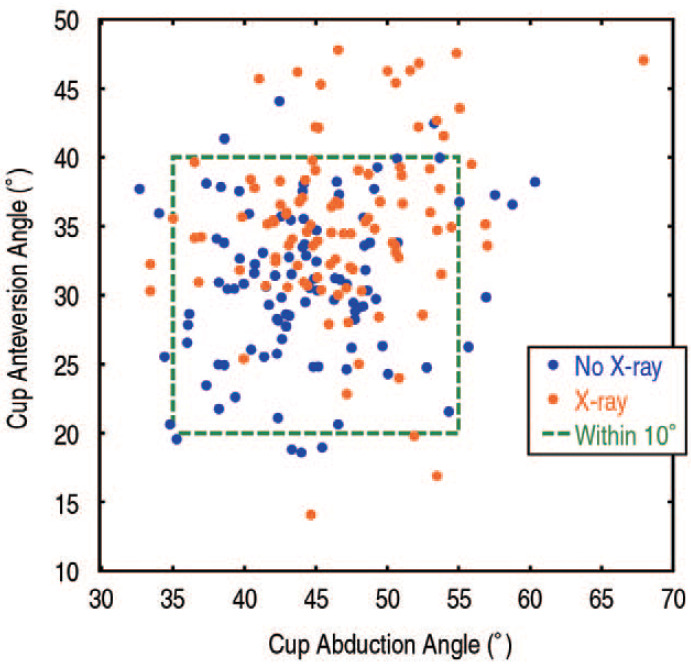
This study evaluated acetabular cup abduction, anteversion, leg length, and offset among 100 posterior approach THAs performed without imaging (No X-ray group) and compared them to a subsequent series of 100 THAs where an intraoperative radiograph was taken with the trial components in place (X-ray group). THAs were performed using a posterior approach by a single, experienced surgeon whose goal was to place the cup at 45° of abduction and 30° of anteversion. Supine anteroposterior pelvic digital radiographs taken at the first (nominal 4-week) postoperative visit were used for measurements.
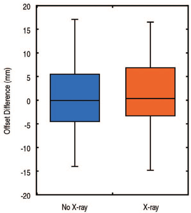
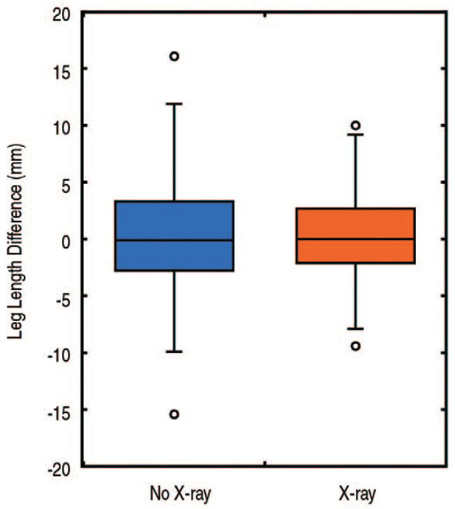
Slight differences in cup abduction (47°±6° vs 44°±6°, respectively, =0.003) and anteversion angle (35°±6° vs 31°±6°, respectively, <0.001) were observed between the X-ray and No X-ray groups; however, a similar proportion of cups within 10° of the target angles was observed (76% vs 83%, respectively, =0.22). No difference in offset measurements (1.1±6.6 mm vs 0.3±6.9 mm, respectively, =0.42) or leg lengths (0.3±3.8 mm vs 0.3±4.8 mm, respectively, =0.94) was observed between the X-ray and No X-ray groups; however, the X-ray group showed less leg length variation (=0.05).
In this study, the routine use of intraoperative radiographs was not associated with improved implant positioning for uncomplicated primary THA.
在全髋关节置换术(THA)手术中,准确的部件放置和患者解剖结构的恢复至关重要。尽管有时会使用术中X线片,但尚不清楚这种做法是否能提高准确性。
本研究评估了100例采用后入路且未使用影像技术的THA(无X线组)的髋臼杯外展、前倾角、腿长和偏移,并将其与随后的100例THA进行比较,后者在试验部件就位后拍摄了术中X线片(X线组)。THA采用后入路,由一位经验丰富的外科医生进行,其目标是将髋臼杯放置在外展45°和前倾角30°的位置。术后首次(名义上的4周)随访时拍摄的仰卧前后位骨盆数字X线片用于测量。
X线组和无X线组之间在髋臼杯外展(分别为47°±6°和44°±6°,P = 0.003)和前倾角(分别为35°±6°和31°±6°,P < 0.001)方面存在轻微差异;然而,观察到目标角度±°范围内的髋臼杯比例相似(分别为76%和83%,P = 0.22)。X线组和无X线组之间在偏移测量(分别为1.1±6.6 mm和0.3±6.9 mm,P = 0.42)或腿长(分别为0.3±3.8 mm和0.3±4.8 mm,P = 0.94)方面未观察到差异;然而,X线组的腿长变化较小(P = 0.05)。
在本研究中,对于无并发症的初次THA,术中X线片的常规使用与改善植入物定位无关。