Discipline of Surgery, Faculty of Health and Medical Sciences, School of Medicine, University of Adelaide, Adelaide, South Australia, Australia.
Department of Surgery, Colorectal Unit, Royal Adelaide Hospital, Adelaide, South Australia, Australia.
BMC Cancer. 2021 Sep 26;21(1):1058. doi: 10.1186/s12885-021-08773-w.
Artificial intelligence (AI) is increasingly being used in medical imaging analysis. We aimed to evaluate the diagnostic accuracy of AI models used for detection of lymph node metastasis on pre-operative staging imaging for colorectal cancer.
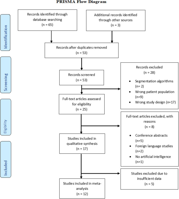
A systematic review was conducted according to PRISMA guidelines using a literature search of PubMed (MEDLINE), EMBASE, IEEE Xplore and the Cochrane Library for studies published from January 2010 to October 2020. Studies reporting on the accuracy of radiomics models and/or deep learning for the detection of lymph node metastasis in colorectal cancer by CT/MRI were included. Conference abstracts and studies reporting accuracy of image segmentation rather than nodal classification were excluded. The quality of the studies was assessed using a modified questionnaire of the QUADAS-2 criteria. Characteristics and diagnostic measures from each study were extracted. Pooling of area under the receiver operating characteristic curve (AUROC) was calculated in a meta-analysis.
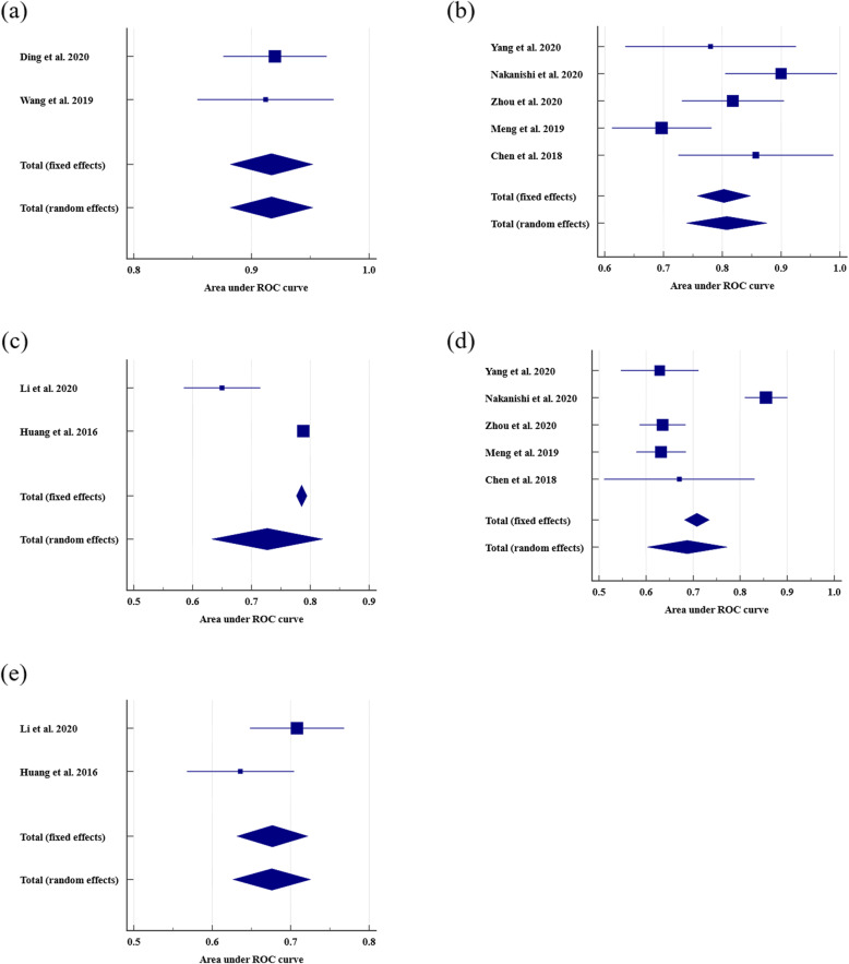
Seventeen eligible studies were identified for inclusion in the systematic review, of which 12 used radiomics models and five used deep learning models. High risk of bias was found in two studies and there was significant heterogeneity among radiomics papers (73.0%). In rectal cancer, there was a per-patient AUROC of 0.808 (0.739-0.876) and 0.917 (0.882-0.952) for radiomics and deep learning models, respectively. Both models performed better than the radiologists who had an AUROC of 0.688 (0.603 to 0.772). Similarly in colorectal cancer, radiomics models with a per-patient AUROC of 0.727 (0.633-0.821) outperformed the radiologist who had an AUROC of 0.676 (0.627-0.725).
AI models have the potential to predict lymph node metastasis more accurately in rectal and colorectal cancer, however, radiomics studies are heterogeneous and deep learning studies are scarce.
PROSPERO CRD42020218004 .
人工智能(AI)在医学影像分析中得到了越来越多的应用。我们旨在评估用于检测结直肠癌术前分期影像中淋巴结转移的 AI 模型的诊断准确性。
根据 PRISMA 指南,我们通过对 PubMed(MEDLINE)、EMBASE、IEEE Xplore 和 Cochrane 图书馆自 2010 年 1 月至 2020 年 10 月发表的研究进行文献检索,进行了系统评价。纳入报告 CT/MRI 用于检测结直肠癌淋巴结转移的放射组学模型和/或深度学习准确性的研究。排除报告图像分割而不是淋巴结分类准确性的会议摘要和研究。使用 QUADAS-2 标准的修改问卷评估研究的质量。从每项研究中提取特征和诊断措施。使用 Meta 分析计算接受者操作特征曲线下面积(AUROC)的汇总。
共确定了 17 项符合纳入标准的系统评价研究,其中 12 项使用了放射组学模型,5 项使用了深度学习模型。两项研究存在高偏倚风险,放射组学论文存在显著异质性(73.0%)。在直肠癌中,放射组学和深度学习模型的每位患者 AUROC 分别为 0.808(0.739-0.876)和 0.917(0.882-0.952)。两种模型的表现均优于 AUROC 为 0.688(0.603-0.772)的放射科医生。同样在结直肠癌中,放射组学模型的每位患者 AUROC 为 0.727(0.633-0.821),优于 AUROC 为 0.676(0.627-0.725)的放射科医生。
AI 模型有可能更准确地预测直肠癌和结直肠癌的淋巴结转移,但放射组学研究存在异质性,深度学习研究稀缺。
PROSPERO CRD42020218004。