Wellman Center for Photomedicine, Massachusetts General Hospital, Boston, Massachusetts 02114, United States.
Harvard Medical School, Boston, Massachusetts 02115, United States.
Nano Lett. 2021 Oct 27;21(20):8595-8601. doi: 10.1021/acs.nanolett.1c02291. Epub 2021 Oct 13.
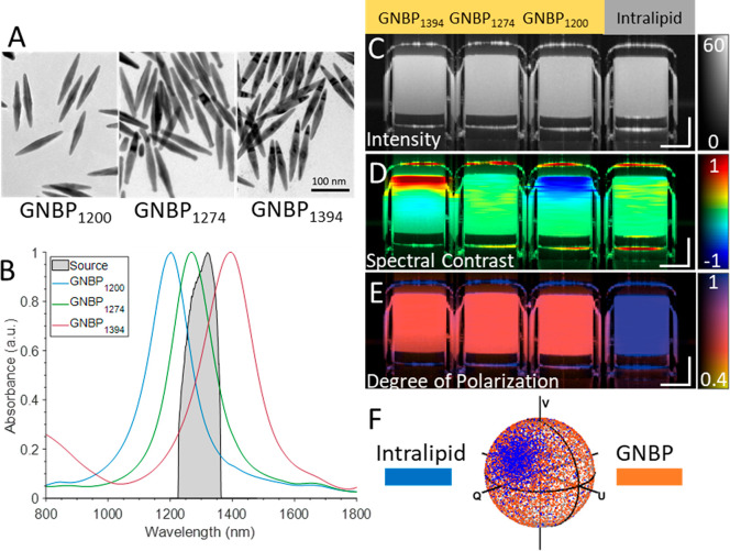
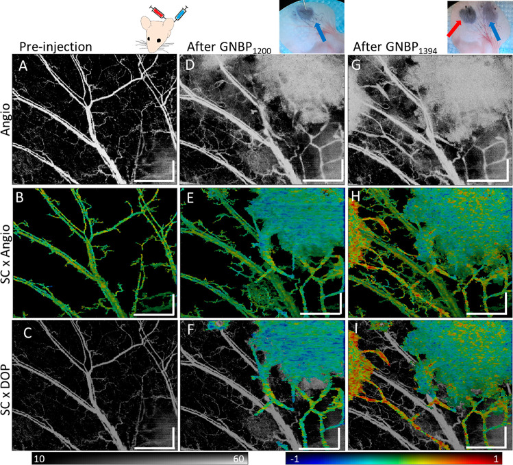
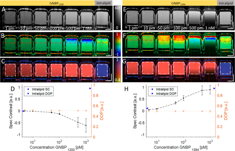
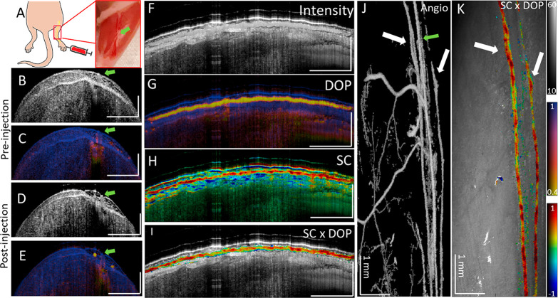
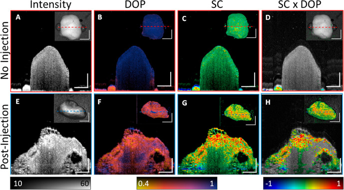
Polarization-sensitive optical coherence tomography (PS-OCT) reveals the subsurface microstructure of biological tissue and provides information regarding the polarization state of light backscattered from tissue. Complementing OCT's structural signal with molecular imaging requires strategies to simultaneously detect multiple exogenous contrast agents with high specificity in tissue. Specific detection of molecular probes enables the parallel visualization of physiological, cellular, and molecular processes. Here we demonstrate that, by combining PS-OCT and spectral contrast (SC)-OCT measurements, we can distinguish signatures of different gold nanobipyramids (GNBPs) in lymphatic vessels from the surrounding tissue and blood vessels in live mouse models. This technique could well be extended to other anisotropic nanoparticle-based OCT contrast agents and presents significant progress toward enabling OCT molecular imaging.
偏振敏感光学相干断层扫描(PS-OCT)揭示了生物组织的亚表面微观结构,并提供了关于组织后向散射光偏振态的信息。为了用分子成像补充 OCT 的结构信号,需要有策略地在组织中同时以高特异性检测多种外源对比剂。分子探针的特异性检测使生理、细胞和分子过程的并行可视化成为可能。在这里,我们证明通过结合 PS-OCT 和光谱对比(SC)-OCT 测量,我们可以区分活小鼠模型中淋巴管中不同金纳米双锥体(GNBPs)的特征与周围组织和血管的特征。该技术很可能扩展到其他各向异性基于纳米颗粒的 OCT 对比剂,并朝着实现 OCT 分子成像的方向取得了重大进展。