• 文献检索
  • 文档翻译
  • 深度研究
  • 学术资讯
  • Suppr Zotero 插件Zotero 插件
  • 邀请有礼
  • 套餐&价格
  • 历史记录
应用&插件
Suppr Zotero 插件Zotero 插件浏览器插件Mac 客户端Windows 客户端微信小程序
定价
高级版会员购买积分包购买API积分包
服务
文献检索文档翻译深度研究API 文档MCP 服务
关于我们
关于 Suppr公司介绍联系我们用户协议隐私条款
关注我们

Suppr 超能文献

核心技术专利:CN118964589B侵权必究
粤ICP备2023148730 号-1Suppr @ 2026

文献检索

告别复杂PubMed语法,用中文像聊天一样搜索,搜遍4000万医学文献。AI智能推荐,让科研检索更轻松。

立即免费搜索

文件翻译

保留排版,准确专业,支持PDF/Word/PPT等文件格式,支持 12+语言互译。

免费翻译文档

深度研究

AI帮你快速写综述,25分钟生成高质量综述,智能提取关键信息,辅助科研写作。

立即免费体验

MesoNet 利用机器学习实现了鼠标中尺度皮质图谱的自动缩放和分割。

MesoNet allows automated scaling and segmentation of mouse mesoscale cortical maps using machine learning.

机构信息

University of British Columbia, Department of Psychiatry, Kinsmen Laboratory of Neurological Research, Detwiller Pavilion, 2255 Wesbrook Mall, Vancouver, V6T 1Z3, British Columbia, Canada.

Department of Psychology, Djavad Mowafaghian Centre for Brain Health, University of British Columbia, Vancouver, British Columbia, Canada.

出版信息

Nat Commun. 2021 Oct 13;12(1):5992. doi: 10.1038/s41467-021-26255-2.

DOI:10.1038/s41467-021-26255-2
PMID:34645817
原文链接:https://pmc.ncbi.nlm.nih.gov/articles/PMC8514445/
Abstract

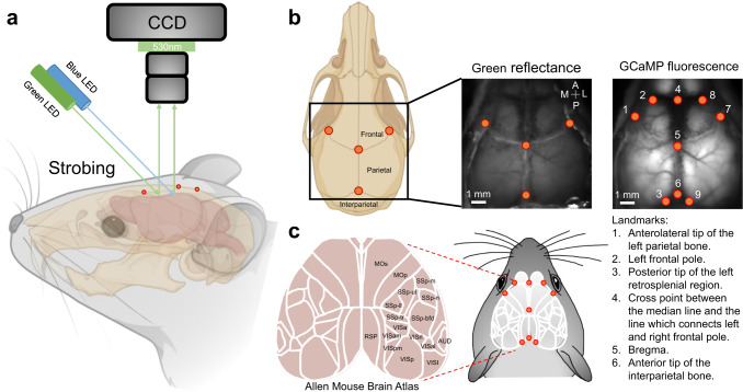
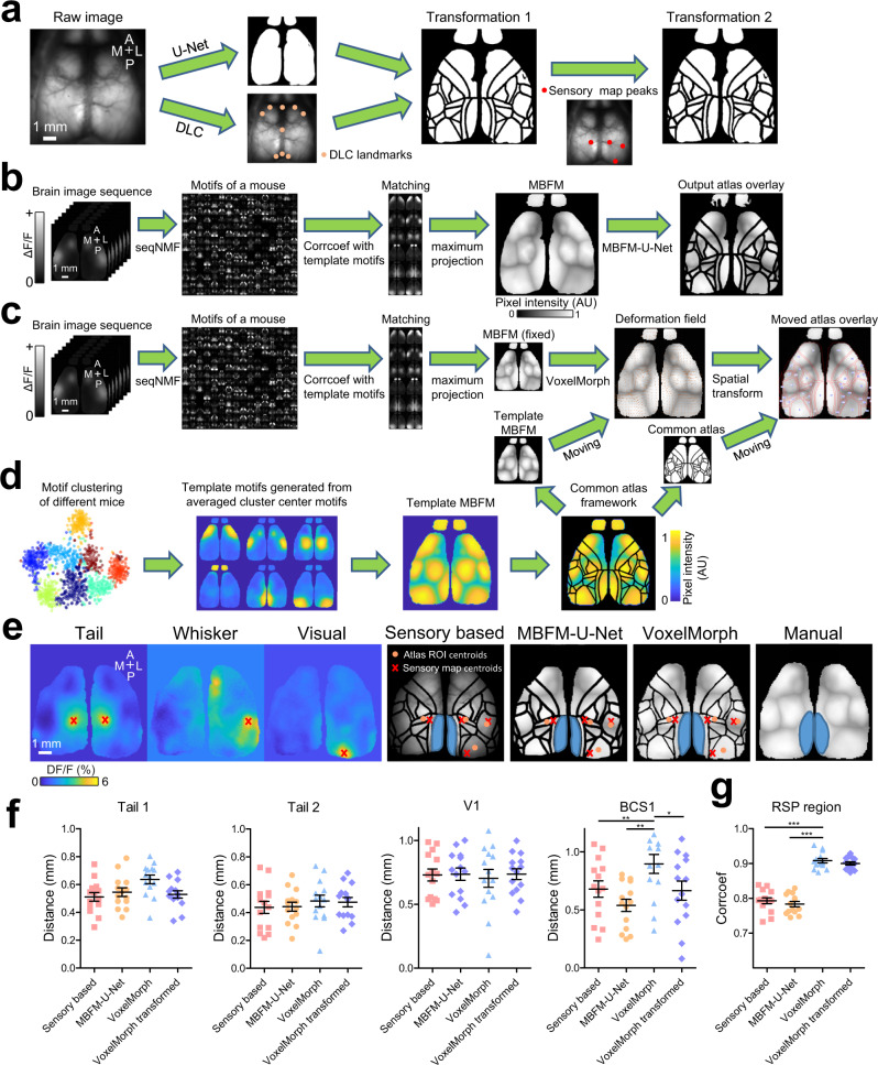
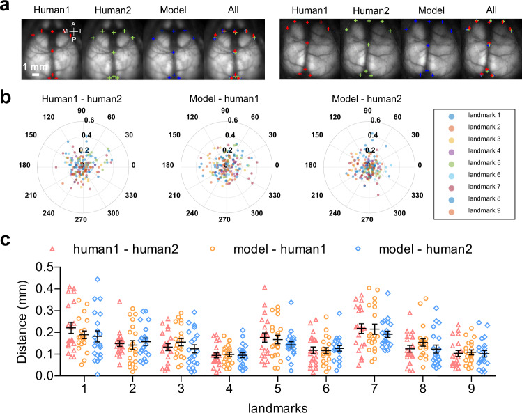
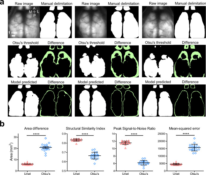
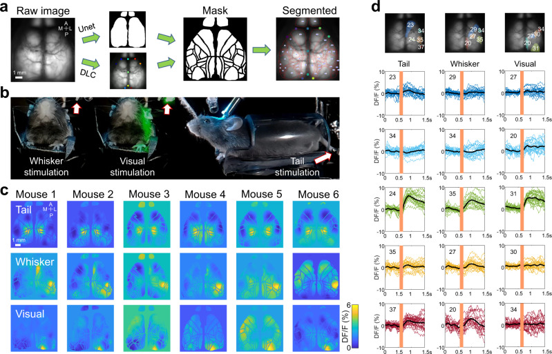
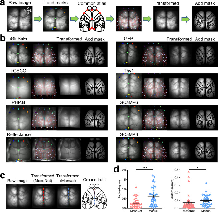
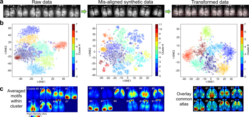
Understanding the basis of brain function requires knowledge of cortical operations over wide spatial scales and the quantitative analysis of brain activity in well-defined brain regions. Matching an anatomical atlas to brain functional data requires substantial labor and expertise. Here, we developed an automated machine learning-based registration and segmentation approach for quantitative analysis of mouse mesoscale cortical images. A deep learning model identifies nine cortical landmarks using only a single raw fluorescent image. Another fully convolutional network was adapted to delimit brain boundaries. This anatomical alignment approach was extended by adding three functional alignment approaches that use sensory maps or spatial-temporal activity motifs. We present this methodology as MesoNet, a robust and user-friendly analysis pipeline using pre-trained models to segment brain regions as defined in the Allen Mouse Brain Atlas. This Python-based toolbox can also be combined with existing methods to facilitate high-throughput data analysis.

摘要

理解大脑功能的基础需要了解广泛空间尺度上的皮质操作,以及在明确定义的大脑区域中对大脑活动进行定量分析。将解剖图谱与大脑功能数据匹配需要大量的工作和专业知识。在这里,我们开发了一种基于自动化机器学习的注册和分割方法,用于对小鼠介观皮质图像进行定量分析。一个深度学习模型仅使用单个原始荧光图像就能识别九个皮质地标。另一个全卷积网络被用于划定大脑边界。通过添加三个使用感觉图谱或时空活动模式的功能对齐方法,扩展了这种解剖配准方法。我们将这种方法称为 MesoNet,这是一种使用预训练模型来分割大脑区域的强大且用户友好的分析管道,这些大脑区域是根据艾伦小鼠脑图谱定义的。这个基于 Python 的工具包还可以与现有的方法结合使用,以方便进行高通量数据分析。

https://cdn.ncbi.nlm.nih.gov/pmc/blobs/4e76/8514445/247d66eb8905/41467_2021_26255_Fig7_HTML.jpg
https://cdn.ncbi.nlm.nih.gov/pmc/blobs/4e76/8514445/36df74762200/41467_2021_26255_Fig1_HTML.jpg
https://cdn.ncbi.nlm.nih.gov/pmc/blobs/4e76/8514445/b2878f791adb/41467_2021_26255_Fig2_HTML.jpg
https://cdn.ncbi.nlm.nih.gov/pmc/blobs/4e76/8514445/3e1ebeb3ab76/41467_2021_26255_Fig3_HTML.jpg
https://cdn.ncbi.nlm.nih.gov/pmc/blobs/4e76/8514445/1bee67821d2d/41467_2021_26255_Fig4_HTML.jpg
https://cdn.ncbi.nlm.nih.gov/pmc/blobs/4e76/8514445/cf49b894d690/41467_2021_26255_Fig5_HTML.jpg
https://cdn.ncbi.nlm.nih.gov/pmc/blobs/4e76/8514445/829963ea1307/41467_2021_26255_Fig6_HTML.jpg
https://cdn.ncbi.nlm.nih.gov/pmc/blobs/4e76/8514445/247d66eb8905/41467_2021_26255_Fig7_HTML.jpg

相似文献

1
MesoNet allows automated scaling and segmentation of mouse mesoscale cortical maps using machine learning.MesoNet 利用机器学习实现了鼠标中尺度皮质图谱的自动缩放和分割。
Nat Commun. 2021 Oct 13;12(1):5992. doi: 10.1038/s41467-021-26255-2.
2
Mapping cortical mesoscopic networks of single spiking cortical or sub-cortical neurons.绘制单个发放动作电位的皮层或皮层下神经元的皮层介观网络。
Elife. 2017 Feb 4;6:e19976. doi: 10.7554/eLife.19976.
3
Optical-flow analysis toolbox for characterization of spatiotemporal dynamics in mesoscale optical imaging of brain activity.用于脑活动介观光学成像的时空动力学特征分析的光流分析工具箱。
Neuroimage. 2017 Jun;153:58-74. doi: 10.1016/j.neuroimage.2017.03.034. Epub 2017 Mar 25.
4
Human brain atlas for automated region of interest selection in quantitative susceptibility mapping: application to determine iron content in deep gray matter structures.人脑图谱用于定量磁化率映射中感兴趣区的自动选择:在确定深部灰质结构铁含量中的应用。
Neuroimage. 2013 Nov 15;82:449-69. doi: 10.1016/j.neuroimage.2013.05.127. Epub 2013 Jun 12.
5
Constructing and optimizing 3D atlases from 2D data with application to the developing mouse brain.从二维数据构建和优化 3D 图谱及其在发育中老鼠大脑中的应用。
Elife. 2021 Feb 11;10:e61408. doi: 10.7554/eLife.61408.
6
A human brain atlas derived via n-cut parcellation of resting-state and task-based fMRI data.通过对静息态和任务态功能磁共振成像数据进行n割法分割得出的人类脑图谱。
Magn Reson Imaging. 2016 Feb;34(2):209-18. doi: 10.1016/j.mri.2015.10.036. Epub 2015 Oct 31.
7
Bi-channel image registration and deep-learning segmentation (BIRDS) for efficient, versatile 3D mapping of mouse brain.双通道图像配准和深度学习分割(BIRDS)用于高效、通用的小鼠大脑 3D 映射。
Elife. 2021 Jan 18;10:e63455. doi: 10.7554/eLife.63455.
8
Using deep learning to segment breast and fibroglandular tissue in MRI volumes.利用深度学习对磁共振成像(MRI)容积中的乳腺和纤维腺组织进行分割。
Med Phys. 2017 Feb;44(2):533-546. doi: 10.1002/mp.12079.
9
Mapping mesoscale axonal projections in the mouse brain using a 3D convolutional network.利用 3D 卷积网络绘制小鼠大脑中的中尺度轴突投射。
Proc Natl Acad Sci U S A. 2020 May 19;117(20):11068-11075. doi: 10.1073/pnas.1918465117. Epub 2020 May 1.
10
Interpreting fMRI data: maps, modules and dimensions.解读功能磁共振成像数据:图谱、模块与维度
Nat Rev Neurosci. 2008 Feb;9(2):123-35. doi: 10.1038/nrn2314.

引用本文的文献

1
Bridging the gap: umIT makes complex imaging data accessible to scientists of all backgrounds.弥合差距:umIT使所有背景的科学家都能获取复杂的成像数据。
Neurophotonics. 2025 Jan;12(Suppl 1):S14616. doi: 10.1117/1.NPh.12.S1.S14616. Epub 2025 Aug 22.
2
Characterization of iGABASnFR2 for mesoscale imaging of intracortical GABA dynamics.用于皮质内GABA动力学中尺度成像的iGABASnFR2的特性表征。
Neurophotonics. 2025 Jul;12(3):035006. doi: 10.1117/1.NPh.12.3.035006. Epub 2025 Aug 13.
3
Multimodal dataset linking wide-field calcium imaging to behavior changes in operant lever-pull task in mice.

本文引用的文献

1
Chronic, cortex-wide imaging of specific cell populations during behavior.在行为过程中对特定细胞群体进行慢性、全皮层范围的成像。
Nat Protoc. 2021 Jul;16(7):3241-3263. doi: 10.1038/s41596-021-00527-z. Epub 2021 Jun 2.
2
Fully autonomous mouse behavioral and optogenetic experiments in home-cage.在家庭环境中进行完全自主的小鼠行为和光遗传学实验。
Elife. 2021 May 4;10:e66112. doi: 10.7554/eLife.66112.
3
Sources of widefield fluorescence from the brain.脑的宽场荧光来源。
将宽视野钙成像与小鼠操作性杠杆拉动任务中的行为变化相联系的多模态数据集。
Sci Data. 2025 Jul 29;12(1):1264. doi: 10.1038/s41597-025-05482-y.
4
Connecto-informatics at the mesoscale: current advances in image processing and analysis for mapping the brain connectivity.中尺度的连接信息学:用于绘制大脑连接的图像处理与分析的当前进展
Brain Inform. 2024 Jun 4;11(1):15. doi: 10.1186/s40708-024-00228-9.
5
A deep learning classification task for brain navigation in rodents using micro-Doppler ultrasound imaging.一项使用微多普勒超声成像在啮齿动物中进行脑导航的深度学习分类任务。
Heliyon. 2024 Mar 6;10(5):e27432. doi: 10.1016/j.heliyon.2024.e27432. eCollection 2024 Mar 15.
6
Traumatic brain injury disrupts state-dependent functional cortical connectivity in a mouse model.创伤性脑损伤破坏了小鼠模型中与状态相关的功能皮质连接。
Cereb Cortex. 2024 Jan 31;34(2). doi: 10.1093/cercor/bhae038.
7
Mesotrode chronic simultaneous mesoscale cortical imaging and subcortical or peripheral nerve spiking activity recording in mice.在小鼠中进行介体型慢性同步介观皮质成像以及皮质下或外周神经尖峰活动记录。
Elife. 2023 Nov 14;12:RP87691. doi: 10.7554/eLife.87691.
8
High fidelity sensory-evoked responses in neocortex after intravenous injection of genetically encoded calcium sensors.静脉注射基因编码钙传感器后新皮层中的高保真感觉诱发电位
Front Neurosci. 2023 May 12;17:1181828. doi: 10.3389/fnins.2023.1181828. eCollection 2023.
9
High-throughput image analysis with deep learning captures heterogeneity and spatial relationships after kidney injury.高通量图像分析与深度学习捕捉肾损伤后的异质性和空间关系。
Sci Rep. 2023 Apr 19;13(1):6361. doi: 10.1038/s41598-023-33433-3.
10
Peripersonal encoding of forelimb proprioception in the mouse somatosensory cortex.小鼠体感皮层中前肢本体感觉的近体空间编码。
Nat Commun. 2023 Apr 12;14(1):1866. doi: 10.1038/s41467-023-37575-w.
Elife. 2020 Nov 6;9:e59841. doi: 10.7554/eLife.59841.
4
A Primer on Motion Capture with Deep Learning: Principles, Pitfalls, and Perspectives.深度学习运动捕捉基础:原理、陷阱与展望。
Neuron. 2020 Oct 14;108(1):44-65. doi: 10.1016/j.neuron.2020.09.017.
5
Revealing architectural order with quantitative label-free imaging and deep learning.利用定量无标记成像和深度学习揭示结构秩序。
Elife. 2020 Jul 27;9:e55502. doi: 10.7554/eLife.55502.
6
Low-Dimensional Spatiotemporal Dynamics Underlie Cortex-wide Neural Activity.低维时空动力学是皮质广泛神经活动的基础。
Curr Biol. 2020 Jul 20;30(14):2665-2680.e8. doi: 10.1016/j.cub.2020.04.090. Epub 2020 May 28.
7
The Allen Mouse Brain Common Coordinate Framework: A 3D Reference Atlas.艾伦鼠脑通用坐标系:一个 3D 参考图谱。
Cell. 2020 May 14;181(4):936-953.e20. doi: 10.1016/j.cell.2020.04.007. Epub 2020 May 7.
8
Localized semi-nonnegative matrix factorization (LocaNMF) of widefield calcium imaging data.广域钙成像数据的局部半非负矩阵分解(LocaNMF)。
PLoS Comput Biol. 2020 Apr 13;16(4):e1007791. doi: 10.1371/journal.pcbi.1007791. eCollection 2020 Apr.
9
Spatiotemporal refinement of signal flow through association cortex during learning.学习过程中通过联合皮层的信号流的时空细化。
Nat Commun. 2020 Apr 8;11(1):1744. doi: 10.1038/s41467-020-15534-z.
10
Comparison between transgenic and AAV-PHP.eB-mediated expression of GCaMP6s using wide-field functional imaging of brain activity.利用脑活动的宽场功能成像比较转基因和AAV-PHP.eB介导的GCaMP6s表达。
Neurophotonics. 2019 Apr;6(2):025014. doi: 10.1117/1.NPh.6.2.025014. Epub 2019 Jun 22.