Division of Rheumatology, Department of Internal Medicine, The Jikei University School of Medicine, 3-25-8 Nishi-shimbashi, Minato-ku, Tokyo, 105-8461, Japan.
Department of Neuroscience, The Jikei University School of Medicine, 3-25-8 Nishi-shimbashi, Minato-ku, Tokyo, Japan.
Arthritis Res Ther. 2021 Oct 29;23(1):273. doi: 10.1186/s13075-021-02657-x.
Central nervous system (CNS)-mediated symptoms, such as fatigue, depression, and hyperalgesia, are common complications among patients with rheumatoid arthritis (RA). However, it remains unclear how the peripheral pathology of RA spreads to the brain. Accumulated evidence showing an association between serum cytokine levels and aberrant CNS function suggests that humoral factors participate in this mechanism. In contrast to the well-known early responses of microglia (CNS-resident immune cells) in the area postrema [AP; a brain region lacking a blood-brain barrier (BBB)] to experimental inflammation, microglial alterations in the AP during chronic inflammation like RA remain unclear. Therefore, to determine whether microglia in the AP can react to persistent autoimmune-arthritis conditions, we analyzed these cells in a mouse model of collagen-induced arthritis (CIA).
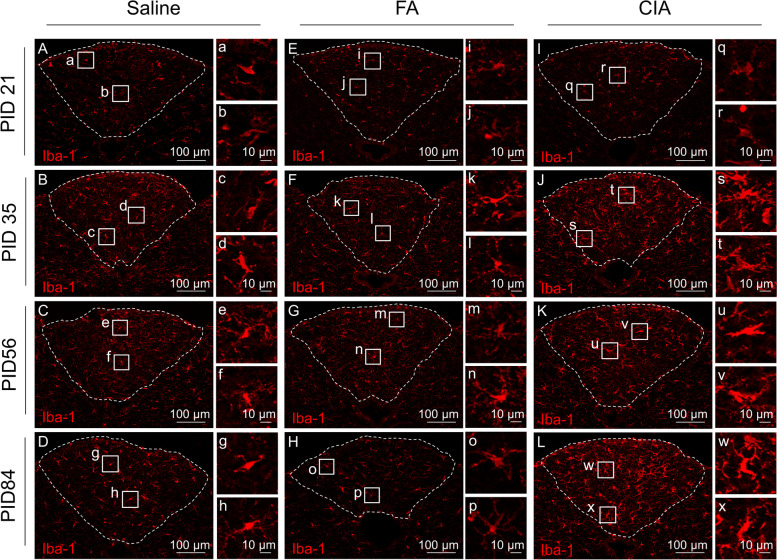
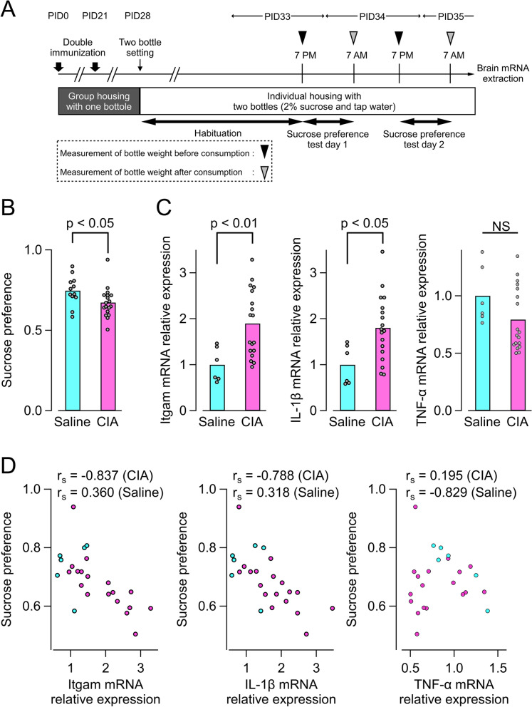
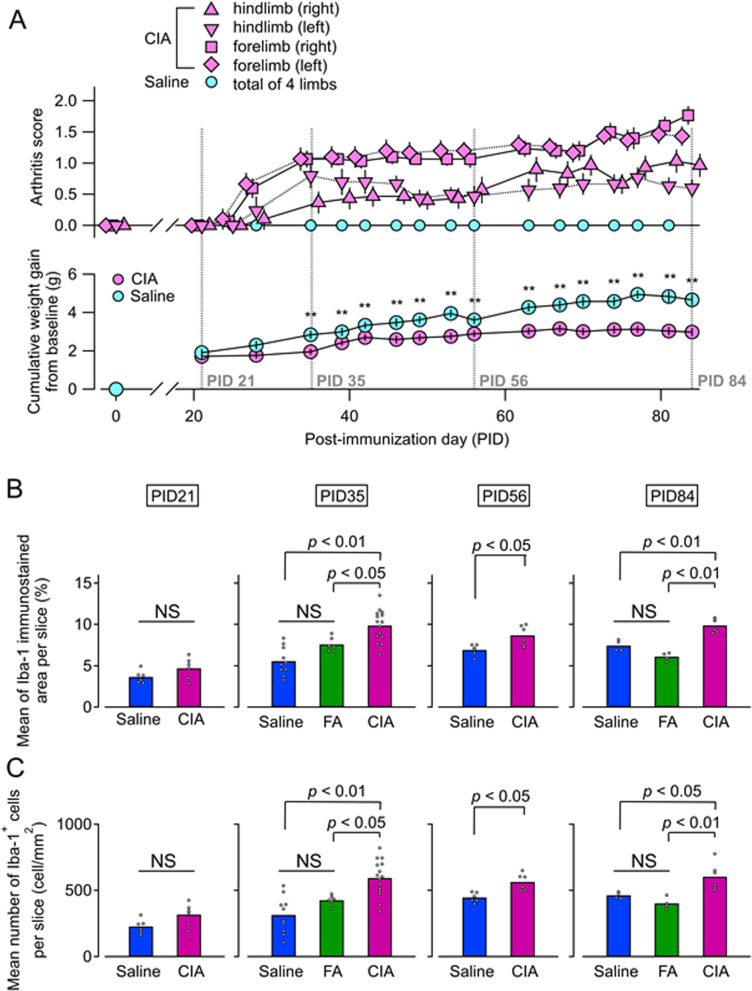
Microglial number and morphology were analyzed in the AP of CIA and control mice (administered Freund's adjuvant or saline). Immunostaining for ionized calcium-binding adaptor molecule-1 was performed at various disease phases: "pre-onset" [post-immunization day (PID) 21], "establishment" (PID 35), and "chronic" (PID 56 and 84). Quantitative analyses of microglial number and morphology were performed, with principal component analysis used to classify microglia. Interleukin-1β (IL-1β) mRNA expression was analyzed by multiple fluorescent in situ hybridization and real-time polymerase chain reaction. Behavioral changes were assessed by sucrose preference test.
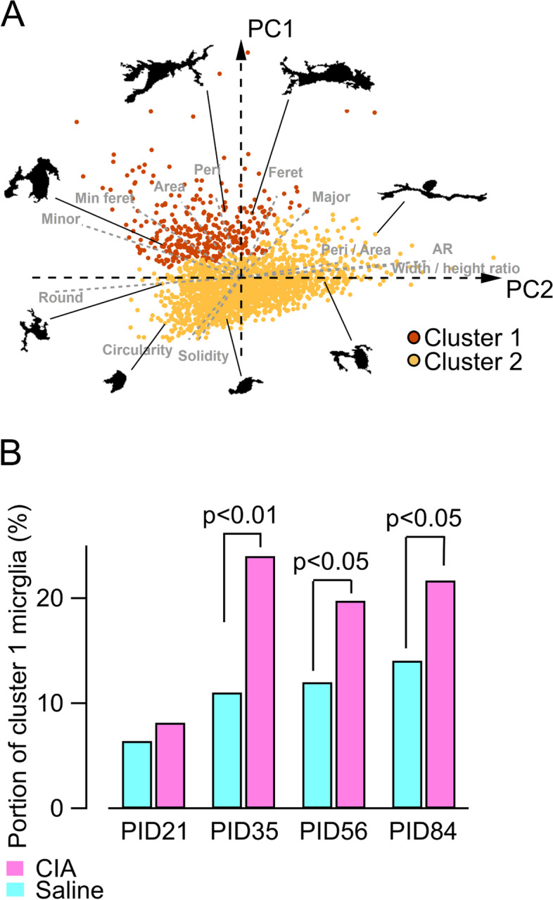
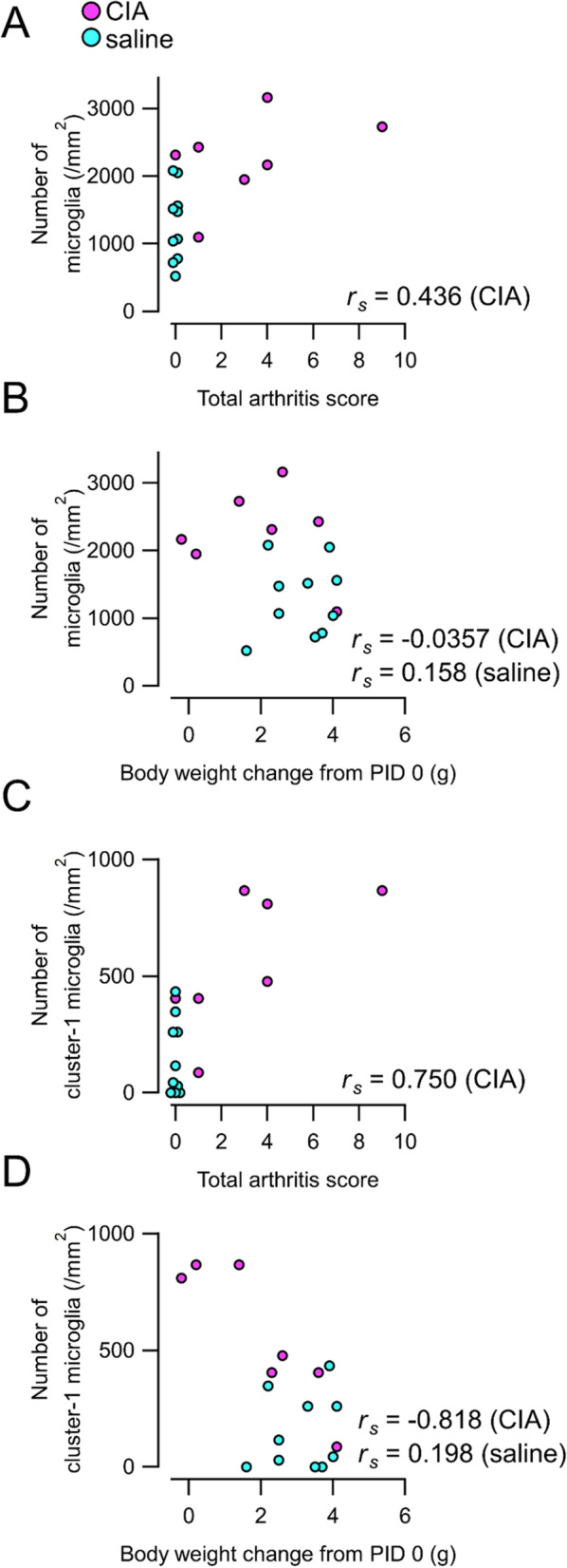
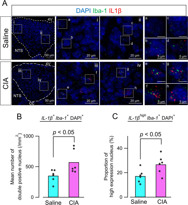
Microglia in the AP significantly increased in density and exhibited changes in morphology during the establishment and chronic phases, but not the pre-onset phase. Non-subjective clustering classification of cell morphology (CIA, 1,256 cells; saline, 852 cells) showed that the proportion of highly activated microglia increased in the CIA group during establishment and chronic phases. Moreover, the density of IL-1β-positive microglia, a hallmark of functional activation, was increased in the AP. Sucrose preferences in CIA mice negatively correlated with IL-1β expression in brain regions containing the AP.
Our findings demonstrate that microglia in the AP can sustain their activated state during persistent autoimmune arthritis, which suggests that chronic inflammation, such as RA, may affect microglia in brain regions lacking a BBB and have various neural consequences.
中枢神经系统(CNS)介导的症状,如疲劳、抑郁和痛觉过敏,是类风湿关节炎(RA)患者的常见并发症。然而,RA 的外周病理学如何扩散到大脑仍不清楚。越来越多的证据表明,血清细胞因子水平与异常的中枢神经系统功能之间存在关联,这表明体液因素参与了这一机制。与实验性炎症中后rema 区[AP;缺乏血脑屏障(BBB)的脑区]中常驻免疫细胞小胶质细胞的早期反应不同,RA 等慢性炎症期间 AP 中小胶质细胞的改变尚不清楚。因此,为了确定 AP 中的小胶质细胞是否能对持续的自身免疫性关节炎状态做出反应,我们在胶原诱导关节炎(CIA)的小鼠模型中分析了这些细胞。
在 CIA 和对照小鼠(给予福氏佐剂或生理盐水)的 AP 中分析小胶质细胞的数量和形态。在疾病的不同阶段进行离子钙结合衔接分子-1的免疫染色:“发作前”[免疫后第 21 天(PID)]、“建立”(PID 35)和“慢性”(PID 56 和 84)。对小胶质细胞数量和形态进行定量分析,并使用主成分分析对小胶质细胞进行分类。通过多重荧光原位杂交和实时聚合酶链反应分析白细胞介素-1β(IL-1β)mRNA 的表达。通过蔗糖偏好试验评估行为变化。
AP 中的小胶质细胞在建立和慢性阶段的密度显著增加,并表现出形态学变化,但在发作前阶段没有变化。细胞形态的非主观聚类分类(CIA,1256 个细胞;生理盐水,852 个细胞)显示,在建立和慢性阶段,CIA 组中高度激活的小胶质细胞比例增加。此外,AP 中功能性激活的标志 IL-1β阳性小胶质细胞的密度增加。CIA 小鼠的蔗糖偏好与 AP 中包含的脑区的 IL-1β表达呈负相关。
我们的研究结果表明,AP 中的小胶质细胞在持续的自身免疫性关节炎中能够维持其激活状态,这表明慢性炎症,如 RA,可能会影响缺乏 BBB 的脑区中的小胶质细胞,并产生各种神经后果。