Department of General Surgery, Zhongshan Hospital, Fudan University, Shanghai, 200032, China.
Cancer Center, Zhongshan Hospital, Fudan University, Shanghai, 200032, China.
Cell Death Dis. 2021 Oct 30;12(11):1033. doi: 10.1038/s41419-021-04293-4.
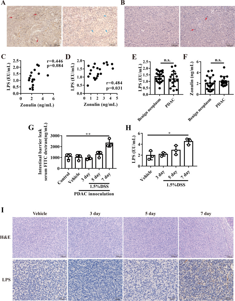
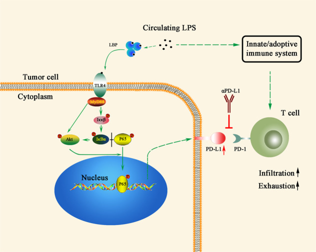
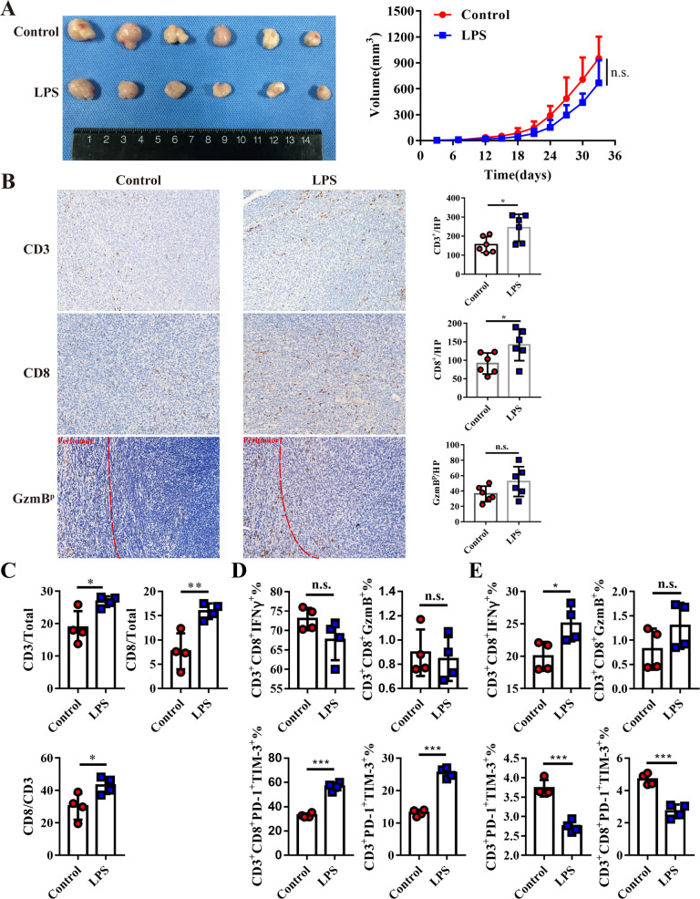
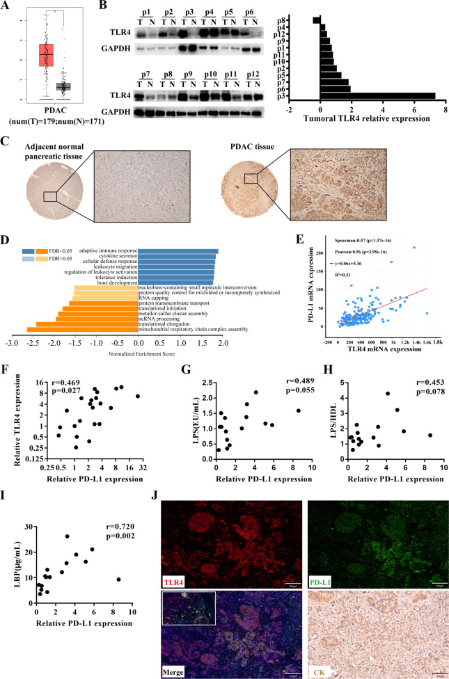
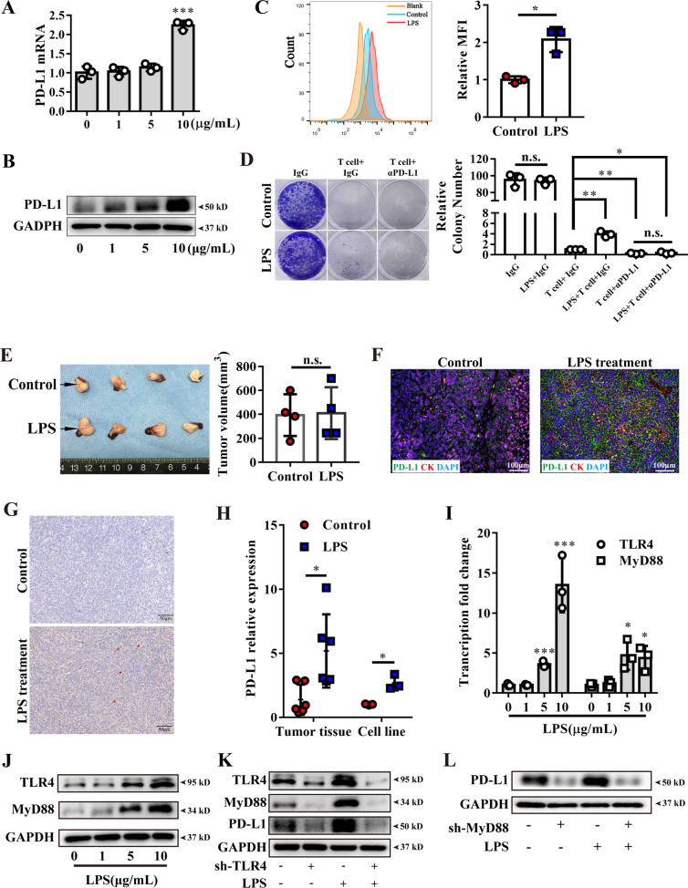
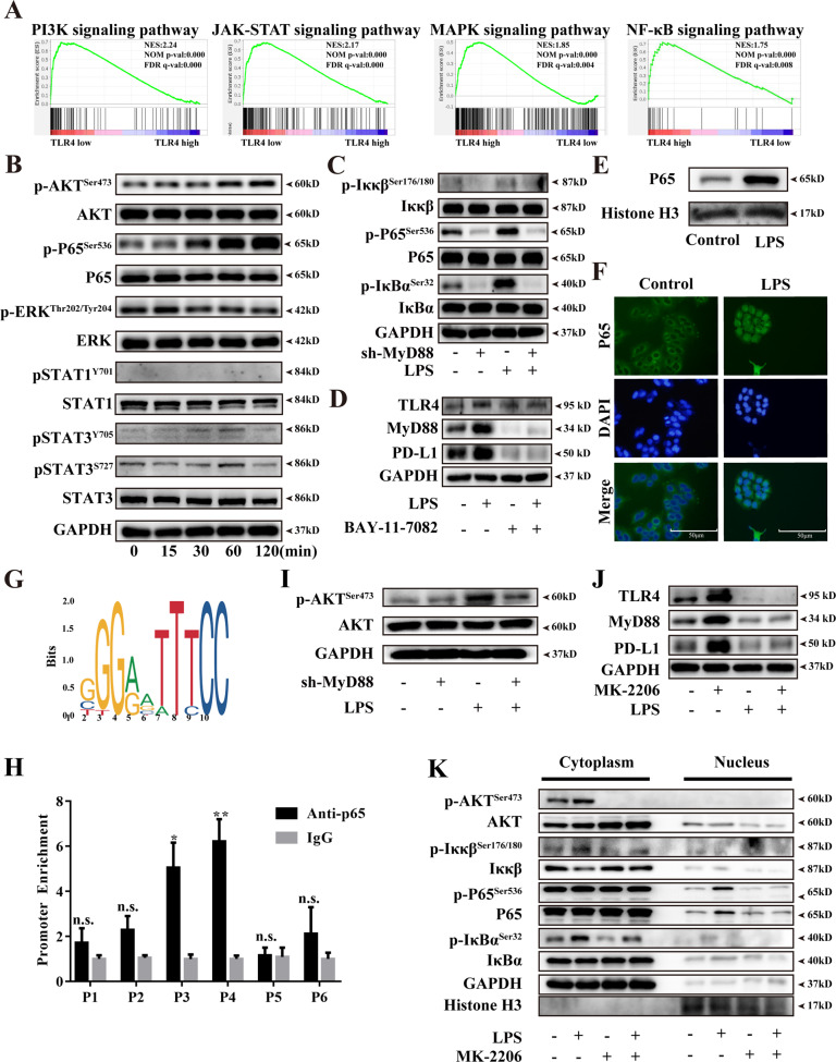
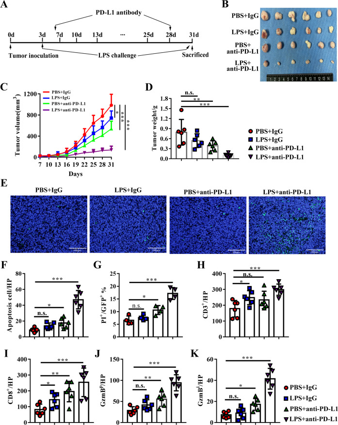
Lipopolysaccharide (LPS) as an important inflammatory mediator activates the innate/adaptive immune system. The existence of LPS in pancreatic ductal adenocarcinoma (PDAC) has been reported, however, its biological function in PDAC remains unclear. Here, we demonstrated that circulating and tumoral LPS was significantly increased by intestinal leakage in the orthotopic murine PDAC model, and LPS administration promoted T cell infiltration but exhaustion paradoxically in the subcutaneous murine PDAC model. By bioinformatic analysis, Toll-like receptor 4 (TLR4), LPS receptor, was further found to enrich in immune tolerance signaling in PDAC tissues. Then, a significant positive correlation was found between TLR4 and programmed death ligand-1 (PD-L1) in clinical PDAC tissues, as well as serum LPS and tumoral PD-L1. Meanwhile, LPS stimulation in vitro and in vivo obviously upregulated tumor PD-L1 expression, and effectively promoted cancer cells resistance to T cell cytotoxicity. Mechanistically, the activation of TLR4/MyD88/AKT/NF-κB cascade was found to participate in LPS mediated PD-L1 transcription via binding to its promoter regions, which was enhanced by crosstalk between NF-κB and AKT pathways. Finally, PD-L1 blockade could significantly reverse LPS-induced immune escape, and synergized with LPS treatment. Taken together, LPS can remodel tumor microenvironment, and synergize with PD-L1 blockade to suppress tumor growth, which may be a promising comprehensive strategy for PDAC.
脂多糖(LPS)作为一种重要的炎症介质,可激活先天/适应性免疫系统。已有报道称胰腺导管腺癌(PDAC)中存在 LPS,但它在 PDAC 中的生物学功能仍不清楚。在这里,我们证明在原位小鼠 PDAC 模型中,肠漏会导致循环和肿瘤 LPS 显著增加,而 LPS 给药可促进皮下小鼠 PDAC 模型中的 T 细胞浸润,但 paradoxically 会导致其衰竭。通过生物信息学分析,进一步发现 Toll 样受体 4(TLR4),即 LPS 受体,在 PDAC 组织中富集于免疫耐受信号中。然后,在临床 PDAC 组织以及血清 LPS 和肿瘤 PD-L1 中发现 TLR4 与程序性死亡配体 1(PD-L1)之间存在显著正相关。同时,LPS 在体外和体内刺激明显上调肿瘤 PD-L1 表达,并有效促进癌细胞对 T 细胞细胞毒性的抵抗力。在机制上,发现 TLR4/MyD88/AKT/NF-κB 级联的激活通过与其启动子区域结合参与 LPS 介导的 PD-L1 转录,该过程受到 NF-κB 和 AKT 通路之间的串扰增强。最后,PD-L1 阻断可显著逆转 LPS 诱导的免疫逃逸,并与 LPS 治疗协同作用。总之,LPS 可以重塑肿瘤微环境,并与 PD-L1 阻断协同抑制肿瘤生长,这可能是一种有前途的 PDAC 综合治疗策略。