Lu Li, Zou Gaocheng, Chen Li, Lu Qianyi, Wu Mian, Li Chunxia
Department of Ophthalmology, The First Affiliated Hospital of University of Science and Technology of China, Hefei, China.
Department of Clinical laboratory, The First Affiliated Hospital of University of Science and Technology of China, Hefei, China.
Front Med (Lausanne). 2021 Oct 15;8:736316. doi: 10.3389/fmed.2021.736316. eCollection 2021.
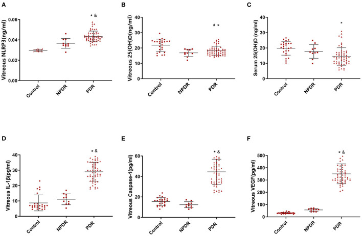
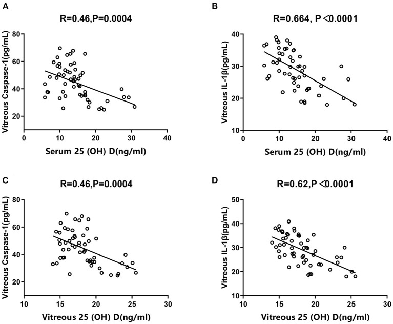
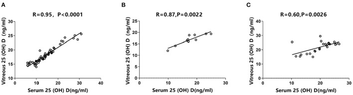
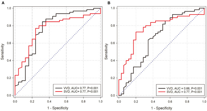
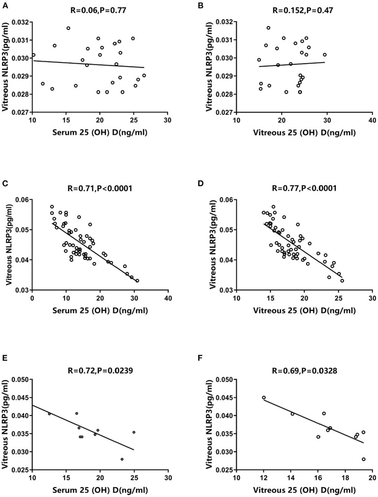
This study aims to determine vitamin D concentrations in the vitreous and serum, as well as the expression levels of NLRP3 inflammasome pathway in the vitreous of patients with proliferative diabetic retinopathy (PDR). In addition, we investigated the possible correlation between NLRP3 inflammasome levels and vitamin D concentrations. We obtained vitreous samples before vitrectomy from 55 PDR patients, 25 non-diabetic patients with idiopathic macular hole (IMH), and 10 non-proliferative diabetic retinopathy (NPDR) patients. We also collected serum samples from the same patients. Enzyme-linked immunosorbent assay (ELISA) was used to examine NLRP3 inflammasome pathway proteins, including NLRP3, caspase-1, IL-1β, and VEGF. In addition, vitamin D concentrations were analyzed in Roche Cobas 6000's module e601 platform using electrochemiluminescence immune assay. The levels of NLRP3 inflammasome pathway and VEGF increased dramatically in PDR vitreous. However, vitamin D concentrations in vitreous and serum followed the opposite trend. Meanwhile, vitreous and serum vitamin D concentrations were significantly negatively correlated with vitreous NLRP3 expression in PDR patients. Moreover, serum and vitreous vitamin D concentrations were positively correlated and demonstrated discriminatory ability in DR. The subgroup analysis of PDR group revealed that eyes with tractional retinal detachment (TRD) had higher NLRP3 inflammasome pathway and VEGF levels but lower vitamin D concentrations. Conversely, eyes that received preoperative pan-retinal photocoagulation (PRP) exhibited lower levels of NLRP3 inflammasome pathway, but vitamin D concentrations were irrelevant to laser treatment. Our results demonstrate a strong correlation between increased NLRP3 inflammasome pathway and decreased vitamin D concentrations in the vitreous of PDR patients, which may be linked to PDR pathogenesis. In addition, vitamin D supplementation may play a key role in preventing, treating, and improving PDR prognosis due to its inhibitory impact on NLRP3 inflammasome pathway and VEGF.
本研究旨在测定增殖性糖尿病视网膜病变(PDR)患者玻璃体和血清中的维生素D浓度,以及玻璃体中NLRP3炎性小体途径的表达水平。此外,我们还研究了NLRP3炎性小体水平与维生素D浓度之间的可能相关性。我们从55例PDR患者、25例非糖尿病特发性黄斑裂孔(IMH)患者和10例非增殖性糖尿病视网膜病变(NPDR)患者中获取了玻璃体切除术前的玻璃体样本。我们还采集了同一批患者的血清样本。采用酶联免疫吸附测定(ELISA)检测NLRP3炎性小体途径蛋白,包括NLRP3、半胱天冬酶-1、白细胞介素-1β和血管内皮生长因子(VEGF)。此外,使用电化学发光免疫测定法在罗氏Cobas 6000的e601模块平台上分析维生素D浓度。PDR玻璃体中NLRP3炎性小体途径和VEGF的水平显著升高。然而,玻璃体和血清中的维生素D浓度呈现相反的趋势。同时,PDR患者玻璃体和血清中的维生素D浓度与玻璃体NLRP3表达显著负相关。此外,血清和玻璃体中的维生素D浓度呈正相关,并在糖尿病视网膜病变中具有鉴别能力。PDR组的亚组分析显示,伴有牵拉性视网膜脱离(TRD)的眼睛具有较高的NLRP3炎性小体途径和VEGF水平,但维生素D浓度较低。相反,接受术前全视网膜光凝(PRP)的眼睛NLRP3炎性小体途径水平较低,但维生素D浓度与激光治疗无关。我们的结果表明,PDR患者玻璃体中NLRP3炎性小体途径增加与维生素D浓度降低之间存在强烈相关性,这可能与PDR的发病机制有关。此外,维生素D补充剂可能因其对NLRP3炎性小体途径和VEGF的抑制作用,在预防、治疗和改善PDR预后方面发挥关键作用。