Elharrouss Omar, Subramanian Nandhini, Al-Maadeed Somaya
Department of Computer Science and Engineering, Qatar University, Doha, Qatar.
SN Comput Sci. 2022;3(1):13. doi: 10.1007/s42979-021-00874-4. Epub 2021 Oct 25.
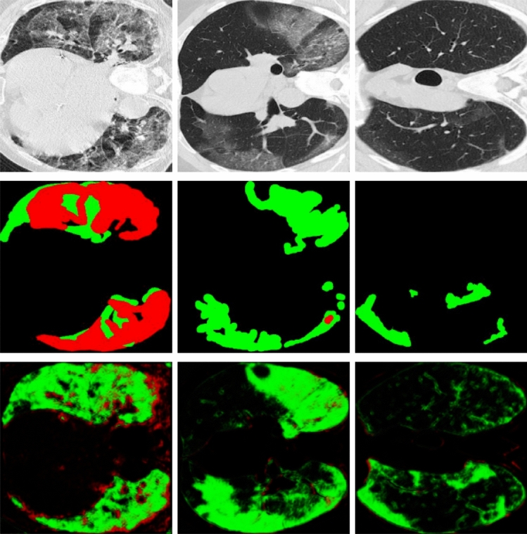
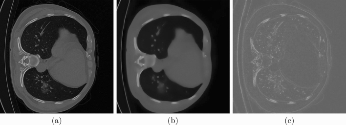
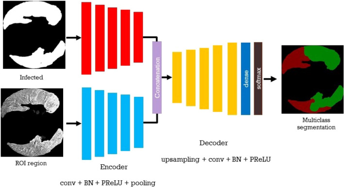
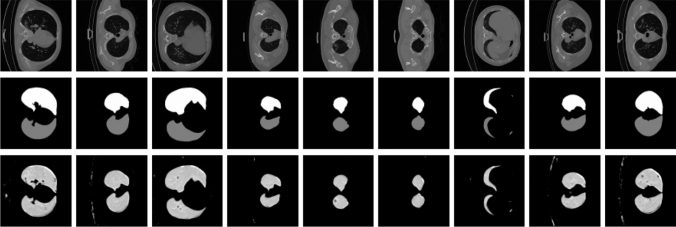
The novelty of the COVID-19 Disease and the speed of spread, created colossal chaotic, impulse all the worldwide researchers to exploit all resources and capabilities to understand and analyze characteristics of the coronavirus in terms of spread ways and virus incubation time. For that, the existing medical features such as CT-scan and X-ray images are used. For example, CT-scan images can be used for the detection of lung infection. However, the quality of these images and infection characteristics limit the effectiveness of these features. Using artificial intelligence (AI) tools and computer vision algorithms, the accuracy of detection can be more accurate and can help to overcome these issues. In this paper, we propose a multi-task deep-learning-based method for lung infection segmentation on CT-scan images. Our proposed method starts by segmenting the lung regions that may be infected. Then, segmenting the infections in these regions. In addition, to perform a multi-class segmentation the proposed model is trained using the two-stream inputs. The multi-task learning used in this paper allows us to overcome the shortage of labeled data. In addition, the multi-input stream allows the model to learn from many features that can improve the results. To evaluate the proposed method, many metrics have been used including Sorensen-Dice similarity, Sensitivity, Specificity, Precision, and MAE metrics. As a result of experiments, the proposed method can segment lung infections with high performance even with the shortage of data and labeled images. In addition, comparing with the state-of-the-art method our method achieves good performance results. For example, the proposed method reached 78..6% for Dice, 71.1% for Sensitivity metric, 99.3% for Specificity 85.6% for Precision, and 0.062 for Mean Average Error metric, which demonstrates the effectiveness of the proposed method for lung infection segmentation.
新型冠状病毒肺炎(COVID-19)疾病的新颖性及其传播速度造成了巨大的混乱,促使全球所有研究人员利用一切资源和能力,从传播方式和病毒潜伏期等方面了解和分析冠状病毒的特征。为此,人们使用了诸如CT扫描和X射线图像等现有的医学特征。例如,CT扫描图像可用于检测肺部感染。然而,这些图像的质量和感染特征限制了这些特征的有效性。使用人工智能(AI)工具和计算机视觉算法,检测的准确性可以更高,并且有助于克服这些问题。在本文中,我们提出了一种基于多任务深度学习的方法,用于对CT扫描图像上的肺部感染进行分割。我们提出的方法首先分割可能被感染的肺部区域。然后,分割这些区域中的感染部位。此外,为了进行多类分割,所提出的模型使用双流输入进行训练。本文中使用的多任务学习使我们能够克服标记数据不足的问题。此外,多输入流允许模型从许多可以改善结果的特征中学习。为了评估所提出的方法,使用了许多指标,包括索伦森-戴斯相似性、灵敏度、特异性、精度和平均绝对误差指标。实验结果表明,即使在数据和标记图像不足的情况下,所提出的方法也能以高性能分割肺部感染。此外,与现有方法相比,我们的方法取得了良好的性能结果。例如,所提出的方法在戴斯指标上达到了78.6%,在灵敏度指标上达到了71.1%,在特异性指标上达到了99.3%,在精度指标上达到了85.6%,在平均绝对误差指标上达到了0.062,这证明了所提出的方法在肺部感染分割方面的有效性。