General Electric Healthcare, Buc, France; LITIS - EA4108 - Quantif, University of Rouen, Rouen, France.
LITIS - EA4108 - Quantif, University of Rouen, Rouen, France; Nuclear Medicine Department, Henri Becquerel Center, Rouen, France.
Comput Biol Med. 2020 Nov;126:104037. doi: 10.1016/j.compbiomed.2020.104037. Epub 2020 Oct 8.
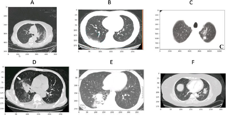
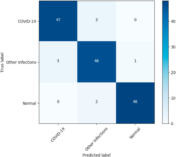
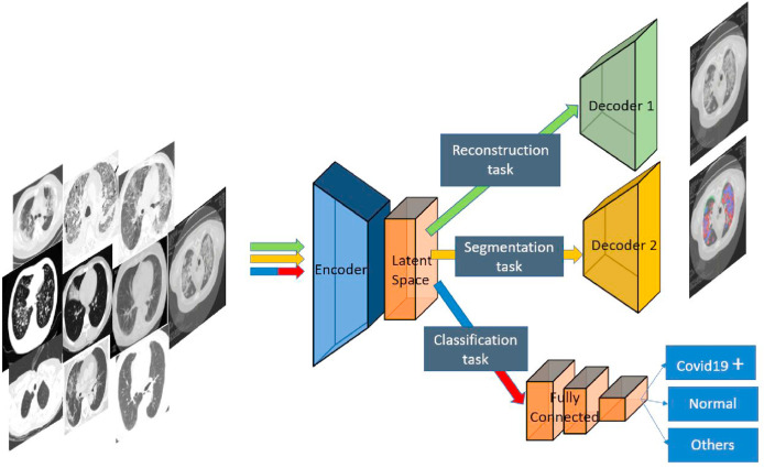
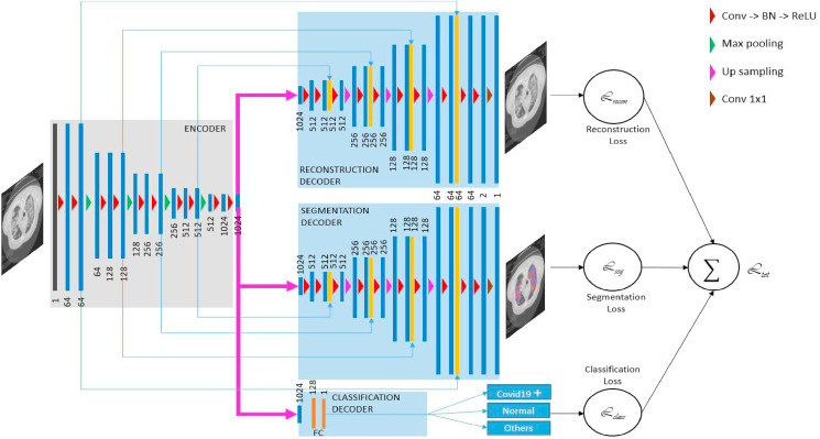
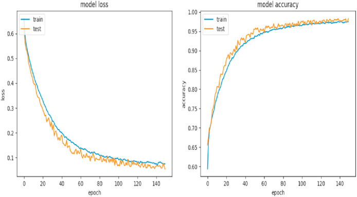
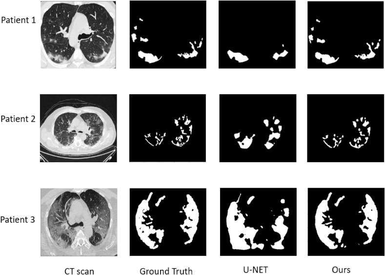
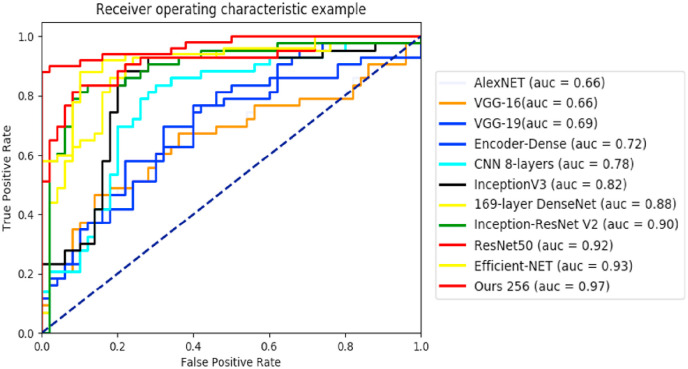
This paper presents an automatic classification segmentation tool for helping screening COVID-19 pneumonia using chest CT imaging. The segmented lesions can help to assess the severity of pneumonia and follow-up the patients. In this work, we propose a new multitask deep learning model to jointly identify COVID-19 patient and segment COVID-19 lesion from chest CT images. Three learning tasks: segmentation, classification and reconstruction are jointly performed with different datasets. Our motivation is on the one hand to leverage useful information contained in multiple related tasks to improve both segmentation and classification performances, and on the other hand to deal with the problems of small data because each task can have a relatively small dataset. Our architecture is composed of a common encoder for disentangled feature representation with three tasks, and two decoders and a multi-layer perceptron for reconstruction, segmentation and classification respectively. The proposed model is evaluated and compared with other image segmentation techniques using a dataset of 1369 patients including 449 patients with COVID-19, 425 normal ones, 98 with lung cancer and 397 of different kinds of pathology. The obtained results show very encouraging performance of our method with a dice coefficient higher than 0.88 for the segmentation and an area under the ROC curve higher than 97% for the classification.
本文提出了一种自动分类分割工具,用于帮助使用胸部 CT 成像筛查 COVID-19 肺炎。分割的病变可以帮助评估肺炎的严重程度并对患者进行随访。在这项工作中,我们提出了一种新的多任务深度学习模型,用于联合识别 COVID-19 患者并从胸部 CT 图像中分割 COVID-19 病变。三个学习任务:分割、分类和重建,使用不同的数据集共同执行。我们的动机一方面是利用多个相关任务中包含的有用信息来提高分割和分类性能,另一方面是处理小数据集的问题,因为每个任务都可以有相对较小的数据集。我们的架构由一个用于解耦特征表示的公共编码器组成,该编码器有三个任务,两个解码器和一个多层感知机,分别用于重建、分割和分类。使用包含 1369 名患者的数据集评估并比较了我们的模型与其他图像分割技术,其中包括 449 名 COVID-19 患者、425 名正常患者、98 名肺癌患者和 397 名不同病理患者。获得的结果表明,我们的方法表现非常出色,分割的骰子系数高于 0.88,分类的 ROC 曲线下面积高于 97%。