• 文献检索
  • 文档翻译
  • 深度研究
  • 学术资讯
  • Suppr Zotero 插件Zotero 插件
  • 邀请有礼
  • 套餐&价格
  • 历史记录
应用&插件
Suppr Zotero 插件Zotero 插件浏览器插件Mac 客户端Windows 客户端微信小程序
定价
高级版会员购买积分包购买API积分包
服务
文献检索文档翻译深度研究API 文档MCP 服务
关于我们
关于 Suppr公司介绍联系我们用户协议隐私条款
关注我们

Suppr 超能文献

核心技术专利:CN118964589B侵权必究
粤ICP备2023148730 号-1Suppr @ 2026

文献检索

告别复杂PubMed语法,用中文像聊天一样搜索,搜遍4000万医学文献。AI智能推荐,让科研检索更轻松。

立即免费搜索

文件翻译

保留排版,准确专业,支持PDF/Word/PPT等文件格式,支持 12+语言互译。

免费翻译文档

深度研究

AI帮你快速写综述,25分钟生成高质量综述,智能提取关键信息,辅助科研写作。

立即免费体验

一种基于肾活检组织学图像的肾小球分类神经网络。

A neural network for glomerulus classification based on histological images of kidney biopsy.

作者信息

Cascarano Giacomo Donato, Debitonto Francesco Saverio, Lemma Ruggero, Brunetti Antonio, Buongiorno Domenico, De Feudis Irio, Guerriero Andrea, Venere Umberto, Matino Silvia, Rocchetti Maria Teresa, Rossini Michele, Pesce Francesco, Gesualdo Loreto, Bevilacqua Vitoantonio

机构信息

Department of Electrical and Information Engineering (DEI), Polytechnic University of Bari, Bary, Italy.

Apulian Bioengineering s.r.l., Modugno, BA, Italy.

出版信息

BMC Med Inform Decis Mak. 2021 Nov 1;21(Suppl 1):300. doi: 10.1186/s12911-021-01650-3.

DOI:10.1186/s12911-021-01650-3
PMID:34724926
原文链接:https://pmc.ncbi.nlm.nih.gov/articles/PMC8559346/
Abstract

BACKGROUND

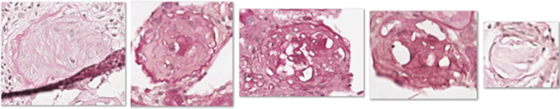
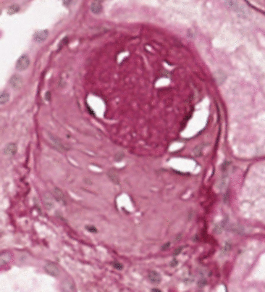
Computer-aided diagnosis (CAD) systems based on medical images could support physicians in the decision-making process. During the last decades, researchers have proposed CAD systems in several medical domains achieving promising results. CAD systems play an important role in digital pathology supporting pathologists in analyzing biopsy slides by means of standardized and objective workflows. In the proposed work, we designed and tested a novel CAD system module based on image processing techniques and machine learning, whose objective was to classify the condition affecting renal corpuscles (glomeruli) between sclerotic and non-sclerotic. Such discrimination is useful for the biopsy slides evaluation performed by pathologists.

RESULTS

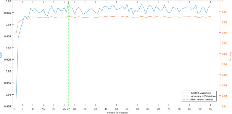
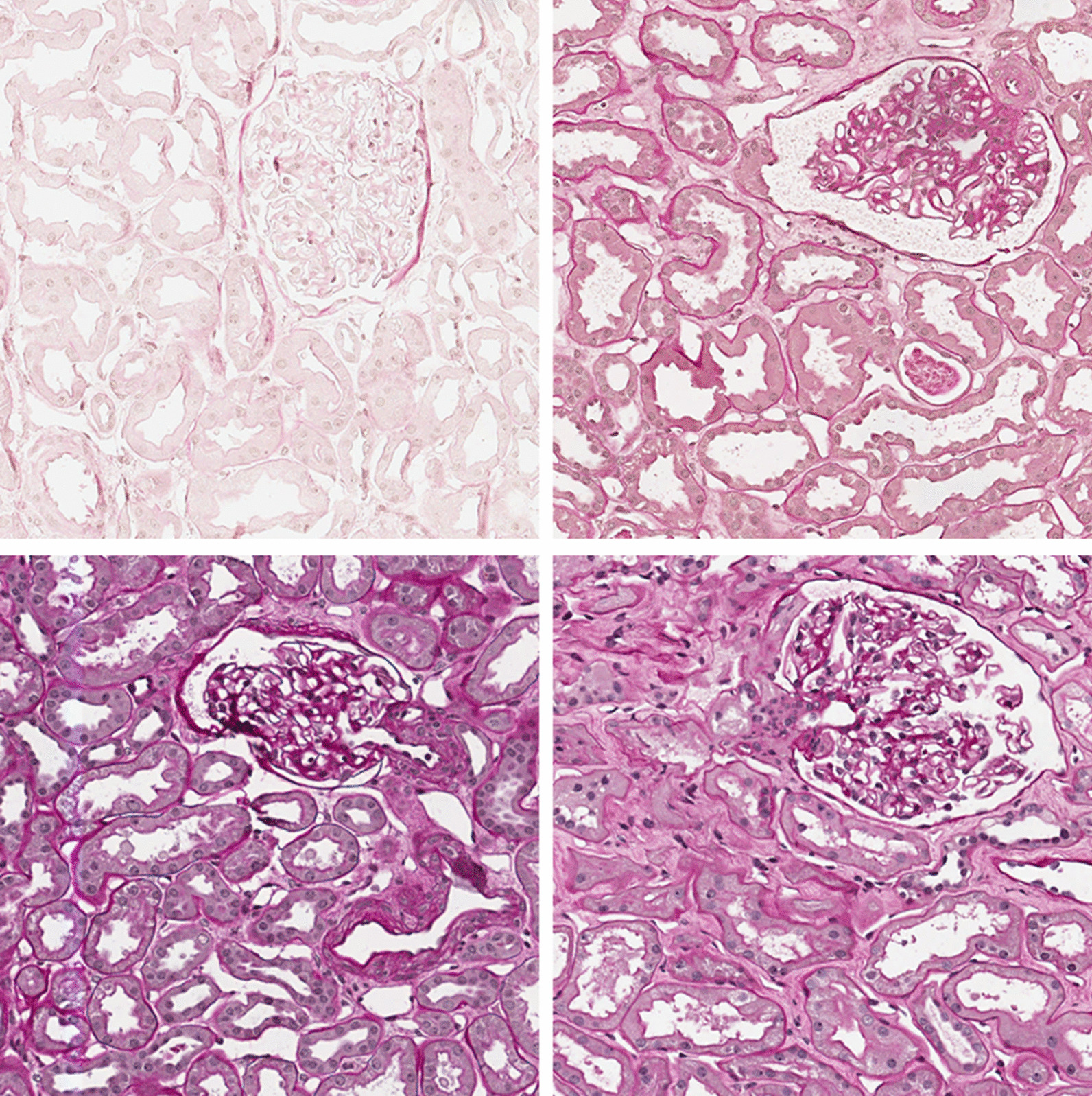
We collected 26 digital slides taken from the kidneys of 19 donors with Periodic Acid-Schiff staining. Expert pathologists have conducted the slides preparation, digital acquisition and glomeruli annotations. Before setting the classifiers, we evaluated several feature extraction techniques from the annotated regions. Then, a feature reduction procedure followed by a shallow artificial neural network allowed discriminating between the glomeruli classes. We evaluated the workflow considering an independent dataset (i.e., processing images not used in the training procedure). Ten independent runs of the training algorithm, and evaluation, allowed achieving MCC and Accuracy of 0.95 (± 0.01) and 0.99 (standard deviation < 0.00), respectively. We also obtained good precision (0.9844 ± 0.0111) and recall (0.9310 ± 0.0153).

CONCLUSIONS

Results on the test set confirm that the proposed workflow is consistent and reliable for the investigated domain, and it can support the clinical practice of discriminating the two classes of glomeruli. Analyses on misclassifications show that the involved images are usually affected by staining artefacts or present partial sections due to slice preparation and staining processes. In clinical practice, however, pathologists discard images showing such artefacts.

摘要

背景

基于医学图像的计算机辅助诊断(CAD)系统可在决策过程中为医生提供支持。在过去几十年中,研究人员已在多个医学领域提出了CAD系统,并取得了有前景的成果。CAD系统在数字病理学中发挥着重要作用,通过标准化和客观的工作流程支持病理学家分析活检切片。在本研究中,我们设计并测试了一种基于图像处理技术和机器学习的新型CAD系统模块,其目的是对影响肾小体(肾小球)的硬化和非硬化状况进行分类。这种区分对病理学家进行活检切片评估很有用。

结果

我们收集了19名捐赠者肾脏的26张经高碘酸-希夫染色的数字切片。专业病理学家进行了切片制备、数字采集和肾小球标注。在设置分类器之前,我们从标注区域评估了几种特征提取技术。然后,经过特征约简过程,再使用浅层人工神经网络对肾小球类别进行区分。我们使用一个独立数据集(即处理训练过程中未使用的图像)评估了该工作流程。训练算法进行十次独立运行和评估后,马修斯相关系数(MCC)和准确率分别达到0.95(±0.01)和0.99(标准差<0.00)。我们还获得了良好的精确率(0.9844±0.0111)和召回率(0.9310±0.0153)。

结论

测试集结果证实,所提出的工作流程对于所研究的领域是一致且可靠的,并且可以支持区分两类肾小球的临床实践。对错误分类的分析表明,所涉及的图像通常受到染色伪影的影响,或者由于切片制备和染色过程而呈现部分切片。然而,在临床实践中,病理学家会丢弃显示此类伪影的图像。

https://cdn.ncbi.nlm.nih.gov/pmc/blobs/0440/8559346/c152c989011c/12911_2021_1650_Fig10_HTML.jpg
https://cdn.ncbi.nlm.nih.gov/pmc/blobs/0440/8559346/a316e13f0ee9/12911_2021_1650_Fig1_HTML.jpg
https://cdn.ncbi.nlm.nih.gov/pmc/blobs/0440/8559346/19cdbc89775b/12911_2021_1650_Fig2_HTML.jpg
https://cdn.ncbi.nlm.nih.gov/pmc/blobs/0440/8559346/0f4e2aca3ee5/12911_2021_1650_Fig3_HTML.jpg
https://cdn.ncbi.nlm.nih.gov/pmc/blobs/0440/8559346/542cc09d5677/12911_2021_1650_Fig4_HTML.jpg
https://cdn.ncbi.nlm.nih.gov/pmc/blobs/0440/8559346/21f884998e11/12911_2021_1650_Fig5_HTML.jpg
https://cdn.ncbi.nlm.nih.gov/pmc/blobs/0440/8559346/5e15652151d6/12911_2021_1650_Fig6_HTML.jpg
https://cdn.ncbi.nlm.nih.gov/pmc/blobs/0440/8559346/8aa7718e5464/12911_2021_1650_Fig7_HTML.jpg
https://cdn.ncbi.nlm.nih.gov/pmc/blobs/0440/8559346/c52239171558/12911_2021_1650_Fig8_HTML.jpg
https://cdn.ncbi.nlm.nih.gov/pmc/blobs/0440/8559346/77d7d1eefacc/12911_2021_1650_Fig9_HTML.jpg
https://cdn.ncbi.nlm.nih.gov/pmc/blobs/0440/8559346/c152c989011c/12911_2021_1650_Fig10_HTML.jpg

相似文献

1
A neural network for glomerulus classification based on histological images of kidney biopsy.一种基于肾活检组织学图像的肾小球分类神经网络。
BMC Med Inform Decis Mak. 2021 Nov 1;21(Suppl 1):300. doi: 10.1186/s12911-021-01650-3.
2
Identification of glomerulosclerosis using IBM Watson and shallow neural networks.利用 IBM Watson 和浅层神经网络识别肾小球硬化。
J Nephrol. 2022 May;35(4):1235-1242. doi: 10.1007/s40620-021-01200-0. Epub 2022 Jan 18.
3
Automated identification of glomeruli and synchronised review of special stains in renal biopsies by machine learning and slide registration: a cross-institutional study.通过机器学习和玻片配准自动识别肾活检中的肾小球并同步审查特殊染色:一项跨机构研究。
Histopathology. 2021 Oct;79(4):499-508. doi: 10.1111/his.14376. Epub 2021 Aug 3.
4
Computer assisted recognition of breast cancer in biopsy images via fusion of nucleus-guided deep convolutional features.通过细胞核引导的深度卷积特征融合实现活检图像中乳腺癌的计算机辅助识别。
Comput Methods Programs Biomed. 2020 Oct;194:105531. doi: 10.1016/j.cmpb.2020.105531. Epub 2020 May 11.
5
Automated quantification of renal interstitial fibrosis for computer-aided diagnosis: A comprehensive tissue structure segmentation method.自动化定量评估肾间质纤维化用于计算机辅助诊断:一种全面的组织结构分割方法。
Comput Methods Programs Biomed. 2018 Mar;155:109-120. doi: 10.1016/j.cmpb.2017.12.004. Epub 2017 Dec 12.
6
Artificial neural networks and pathologists recognize basal cell carcinomas based on different histological patterns.人工神经网络和病理学家基于不同的组织学模式识别基底细胞癌。
Mod Pathol. 2021 May;34(5):895-903. doi: 10.1038/s41379-020-00712-7. Epub 2020 Nov 13.
7
A hybrid fuzzy-neural system for computer-aided diagnosis of ultrasound kidney images using prominent features.一种利用显著特征的用于超声肾脏图像计算机辅助诊断的混合模糊神经网络系统。
J Med Syst. 2008 Feb;32(1):65-83. doi: 10.1007/s10916-007-9109-5.
8
Development and Validation of a Deep Learning Model to Quantify Glomerulosclerosis in Kidney Biopsy Specimens.深度学习模型在肾脏活检标本肾小球硬化定量分析中的建立和验证
JAMA Netw Open. 2021 Jan 4;4(1):e2030939. doi: 10.1001/jamanetworkopen.2020.30939.
9
Breast cancer detection from biopsy images using nucleus guided transfer learning and belief based fusion.基于核引导迁移学习和置信度融合的活检图像乳腺癌检测
Comput Biol Med. 2020 Sep;124:103954. doi: 10.1016/j.compbiomed.2020.103954. Epub 2020 Aug 4.
10
Deep Learning Global Glomerulosclerosis in Transplant Kidney Frozen Sections.深度学习移植肾冷冻切片中的全球肾小球硬化症。
IEEE Trans Med Imaging. 2018 Dec;37(12):2718-2728. doi: 10.1109/TMI.2018.2851150. Epub 2018 Jun 27.

引用本文的文献

1
Vision transformer introduces a new vitality to the classification of renal pathology.视觉转换器为肾病理学分类带来了新的活力。
BMC Nephrol. 2024 Oct 9;25(1):337. doi: 10.1186/s12882-024-03800-x.
2
Towards accurate and efficient diagnoses in nephropathology: An AI-based approach for assessing kidney transplant rejection.迈向肾病理学准确高效诊断:一种基于人工智能的评估肾移植排斥反应的方法。
Comput Struct Biotechnol J. 2024 Aug 16;24:571-582. doi: 10.1016/j.csbj.2024.08.011. eCollection 2024 Dec.
3
Artificial Intelligence-Driven Analysis of Antimicrobial-Resistant and Biofilm-Forming Pathogens on Biotic and Abiotic Surfaces.

本文引用的文献

1
Region-Based Convolutional Neural Nets for Localization of Glomeruli in Trichrome-Stained Whole Kidney Sections.基于区域的卷积神经网络在三色染色全肾切片中肾小球定位的应用。
J Am Soc Nephrol. 2018 Aug;29(8):2081-2088. doi: 10.1681/ASN.2017111210. Epub 2018 Jun 19.
2
Multi-radial LBP Features as a Tool for Rapid Glomerular Detection and Assessment in Whole Slide Histopathology Images.多径向 LBP 特征作为一种在全切片病理图像中快速肾小球检测和评估的工具。
Sci Rep. 2018 Feb 1;8(1):2032. doi: 10.1038/s41598-018-20453-7.
3
Ten quick tips for machine learning in computational biology.
人工智能驱动的生物和非生物表面上抗微生物和生物膜形成病原体的分析
Antibiotics (Basel). 2024 Aug 22;13(8):788. doi: 10.3390/antibiotics13080788.
4
Automatic Identification of Glomerular in Whole-Slide Images Using a Modified UNet Model.使用改进的UNet模型自动识别全切片图像中的肾小球
Diagnostics (Basel). 2023 Oct 9;13(19):3152. doi: 10.3390/diagnostics13193152.
5
European Society for Organ Transplantation (ESOT)-TLJ 3.0 Consensus on Histopathological Analysis of Pre-Implantation Donor Kidney Biopsy: Redefining the Role in the Process of Graft Assessment.欧洲器官移植学会(ESOT)-TLJ 3.0 关于移植前供体肾活检的组织病理学分析共识:重新定义在移植物评估过程中的作用。
Transpl Int. 2023 Jul 4;36:11410. doi: 10.3389/ti.2023.11410. eCollection 2023.
6
Artificial Intelligence-Assisted Renal Pathology: Advances and Prospects.人工智能辅助肾病理学:进展与展望
J Clin Med. 2022 Aug 22;11(16):4918. doi: 10.3390/jcm11164918.
计算生物学中机器学习的十条快速提示。
BioData Min. 2017 Dec 8;10:35. doi: 10.1186/s13040-017-0155-3. eCollection 2017.
4
Chronic kidney disease and the global NCDs agenda.慢性肾脏病与全球非传染性疾病议程。
BMJ Glob Health. 2017 Jul 6;2(2):e000380. doi: 10.1136/bmjgh-2017-000380. eCollection 2017.
5
Global, regional, and national life expectancy, all-cause mortality, and cause-specific mortality for 249 causes of death, 1980-2015: a systematic analysis for the Global Burden of Disease Study 2015.1980 - 2015年全球、区域和国家249种死因的预期寿命、全死因死亡率和死因别死亡率:全球疾病负担研究2015的系统分析
Lancet. 2016 Oct 8;388(10053):1459-1544. doi: 10.1016/S0140-6736(16)31012-1.
6
Measurement of glomerulus diameter and Bowman's space width of renal albino rats.测量白化病大鼠肾小球直径和鲍曼氏囊腔宽度。
Comput Methods Programs Biomed. 2016 Apr;126:143-53. doi: 10.1016/j.cmpb.2015.10.023. Epub 2015 Dec 19.
7
Segmental HOG: new descriptor for glomerulus detection in kidney microscopy image.分段方向梯度直方图:用于肾脏显微镜图像中肾小球检测的新描述符。
BMC Bioinformatics. 2015 Sep 30;16:316. doi: 10.1186/s12859-015-0739-1.
8
Worldwide access to treatment for end-stage kidney disease: a systematic review.全球范围内终末期肾病治疗的可及性:一项系统评价。
Lancet. 2015 May 16;385(9981):1975-82. doi: 10.1016/S0140-6736(14)61601-9. Epub 2015 Mar 13.
9
ROC operating point selection for classification of imbalanced data with application to computer-aided polyp detection in CT colonography.利用 ROC 操作点选择对不平衡数据进行分类,应用于 CT 结肠成像中的计算机辅助息肉检测。
Int J Comput Assist Radiol Surg. 2014 Jan;9(1):79-89. doi: 10.1007/s11548-013-0913-8.
10
Active contours without edges.无边缘活动轮廓。
IEEE Trans Image Process. 2001;10(2):266-77. doi: 10.1109/83.902291.