Fakultät für Physik, Nano Institute Munich & Center for NanoScience (CeNS), Ludwig-Maximilians-Universität, Königinstr. 10, 80539, Munich, Germany.
Attocube Systems AG, Eglfinger Weg 2, 85540, Haar, Germany.
Sci Rep. 2021 Nov 8;11(1):21860. doi: 10.1038/s41598-021-01425-w.
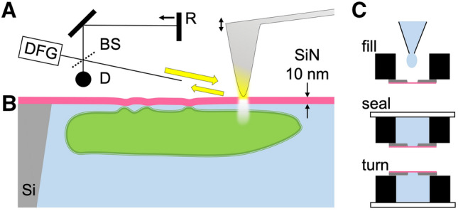
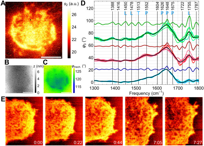
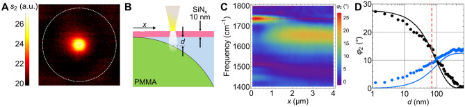
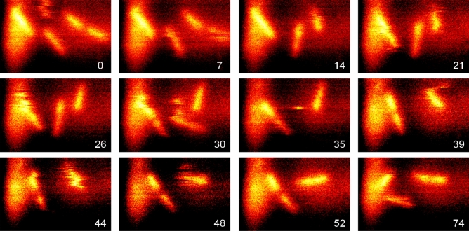
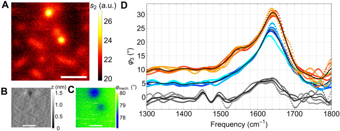
Infrared fingerprint spectra can reveal the chemical nature of materials down to 20-nm detail, far below the diffraction limit, when probed by scattering-type scanning near-field optical microscopy (s-SNOM). But this was impossible with living cells or aqueous processes as in corrosion, due to water-related absorption and tip contamination. Here, we demonstrate infrared s-SNOM of water-suspended objects by probing them through a 10-nm thick SiN membrane. This separator stretches freely over up to 250 µm, providing an upper, stable surface to the scanning tip, while its lower surface is in contact with the liquid and localises adhering objects. We present its proof-of-principle applicability in biology by observing simply drop-casted, living E. coli in nutrient medium, as well as living A549 cancer cells, as they divide, move and develop rich sub-cellular morphology and adhesion patterns, at 150 nm resolution. Their infrared spectra reveal the local abundances of water, proteins, and lipids within a depth of ca. 100 nm below the SiN membrane, as we verify by analysing well-defined, suspended polymer spheres and through model calculations. SiN-membrane based s-SNOM thus establishes a novel tool of live cell nano-imaging that returns structure, dynamics and chemical composition. This method should benefit the nanoscale analysis of any aqueous system, from physics to medicine.
红外指纹光谱可以揭示材料的化学性质,在散射型近场光学显微镜 (s-SNOM) 探测下,其细节可达到 20nm 以下,远低于衍射极限。但由于与水有关的吸收和尖端污染,对于活细胞或水相过程(如腐蚀),这是不可能的。在这里,我们通过探测通过 10nm 厚的 SiN 膜来演示水悬浮物体的红外 s-SNOM。这种分离器可以自由拉伸至 250µm 以上,为扫描尖端提供一个稳定的上表面,而其下表面与液体接触并定位附着的物体。我们通过观察简单地滴铸在营养培养基中的活大肠杆菌以及分裂、移动和发育出丰富的亚细胞形态和粘附模式的活 A549 癌细胞,证明了其在生物学中的原理适用性,分辨率为 150nm。它们的红外光谱揭示了 SiN 膜下方约 100nm 深度处水、蛋白质和脂质的局部丰度,我们通过分析定义明确的悬浮聚合物球和通过模型计算来验证这一点。基于 SiN 膜的 s-SNOM 因此建立了一种新的活细胞纳米成像工具,可提供结构、动态和化学成分。这种方法应该有益于从物理学到医学的任何水相系统的纳米尺度分析。