Korkmaz İnan, Keleş Fatma
Department of Radiology, Hatay Mustafa Kemal University, Faculty of Medicine, Antakya, TUR.
Cureus. 2021 Oct 6;13(10):e18554. doi: 10.7759/cureus.18554. eCollection 2021 Oct.
This study aimed to evaluate the frequency of typical and atypical thoracic CT findings in patient groups diagnosed during different periods of the pandemic, examine disease severity using radiological scoring methods, and determine the relationship between atypical CT findings and disease severity.
One hundred fifty-one patients with positive reverse transcription polymerase chain reaction (RT-PCR) test and thoracic CT scan were included in the study. The patients were divided into two groups as group 1 (March to August 2020) diagnosed in the first six months of the pandemic and group 2 (September 2020 to February 2021) diagnosed in the second six months. CT images of the patients were analyzed for the frequency of typical and atypical findings. Evaluation was made in terms of disease suspicion and severity by scoring methods, and the relationship between atypical findings and disease severity was examined.
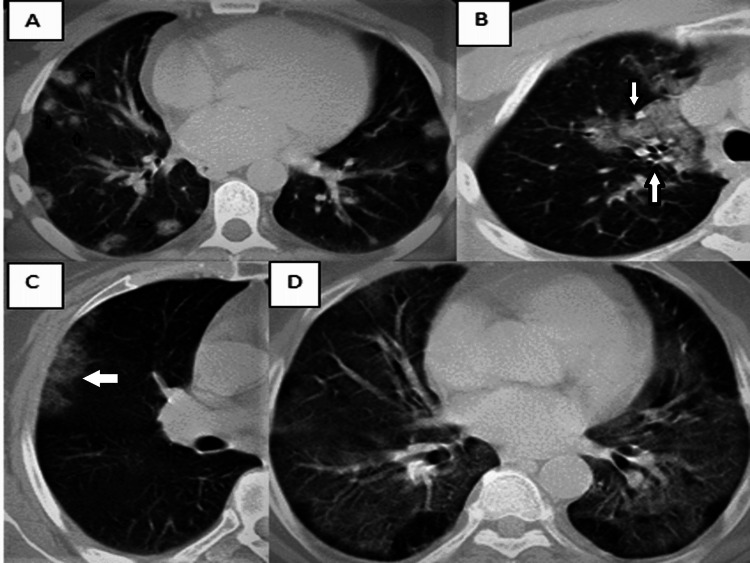
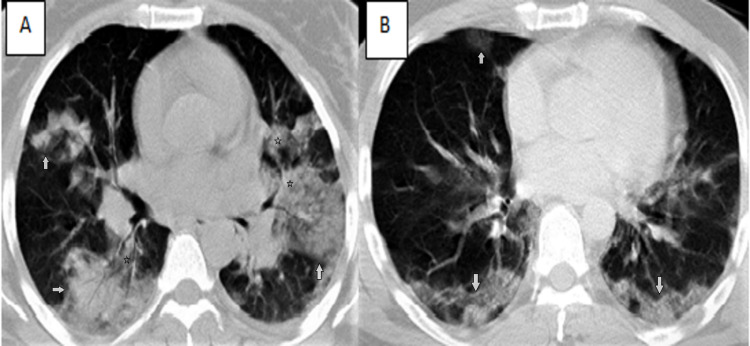
There was no statistically significant difference between the frequency and distribution patterns of typical CT findings observed in both groups. The most common atypical finding in both groups was nodular lesions. Central distribution, one of the atypical findings, was not seen in group 1, whereas it was present in nine patients in group 2 (p=0.001). The mean CT severity score was higher in group 2, and there was a statistically significant difference between the mean CT scores of both groups (p<0.001). In addition, six (7.2%) patients in group 1 and 34 (50%) patients in group 2 had CT scores above the cut-off value (p<0.001). There was no statistically significant relationship between atypical findings and severity score.
Other diseases and atypical findings that may accompany COVID-19 pneumonia may increase the rate of misdiagnosis. In the diagnosis of the disease, clinical signs and symptoms and radiological findings should be evaluated together, and it should be kept in mind that lung findings in thorax CT change over time.
本研究旨在评估在疫情不同时期确诊的患者群体中典型和非典型胸部CT表现的频率,使用放射学评分方法检查疾病严重程度,并确定非典型CT表现与疾病严重程度之间的关系。
本研究纳入了151例逆转录聚合酶链反应(RT-PCR)检测呈阳性且进行了胸部CT扫描的患者。患者被分为两组,第1组(2020年3月至8月)为疫情前六个月确诊的患者,第2组(2020年9月至2021年2月)为后六个月确诊的患者。分析患者的CT图像,以确定典型和非典型表现的频率。通过评分方法对疾病的疑似程度和严重程度进行评估,并检查非典型表现与疾病严重程度之间的关系。
两组观察到的典型CT表现的频率和分布模式之间没有统计学上的显著差异。两组中最常见的非典型表现是结节状病变。非典型表现之一的中央分布在第1组中未观察到,而在第2组中有9例患者出现(p=0.001)。第2组的平均CT严重程度评分更高,两组的平均CT评分之间存在统计学上的显著差异(p<0.001)。此外,第1组中有6例(7.2%)患者和第2组中有34例(50%)患者的CT评分高于临界值(p<0.001)。非典型表现与严重程度评分之间没有统计学上的显著关系。
COVID-19肺炎可能伴随的其他疾病和非典型表现可能会增加误诊率。在疾病诊断中,应综合评估临床体征和症状以及放射学表现,并应牢记胸部CT中的肺部表现会随时间变化。