Polymers and Complex Fluids Group, National Institute of Standards and Technology, 100 Bureau Drive, Gaithersburg, MD, USA.
ADA Science and Research Institute, 100 Bureau Dr, Gaithersburg, MD, 20899, USA.
Soft Matter. 2021 Dec 22;18(1):117-125. doi: 10.1039/d1sm01312b.
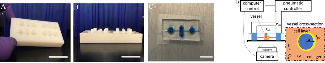
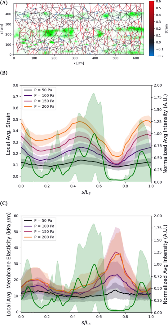
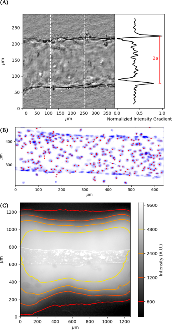
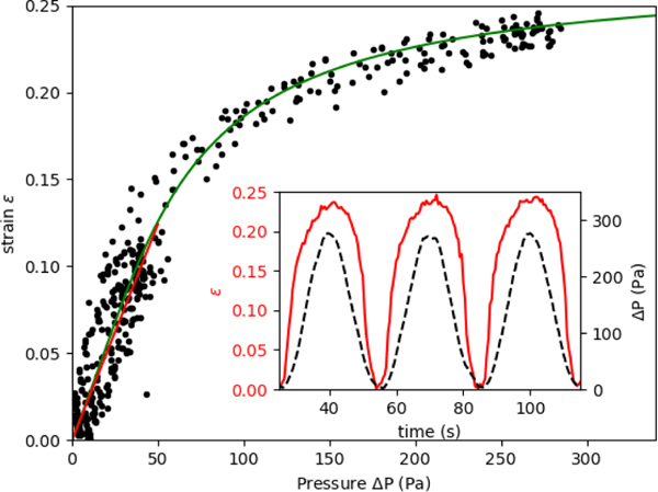
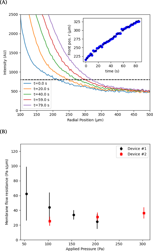
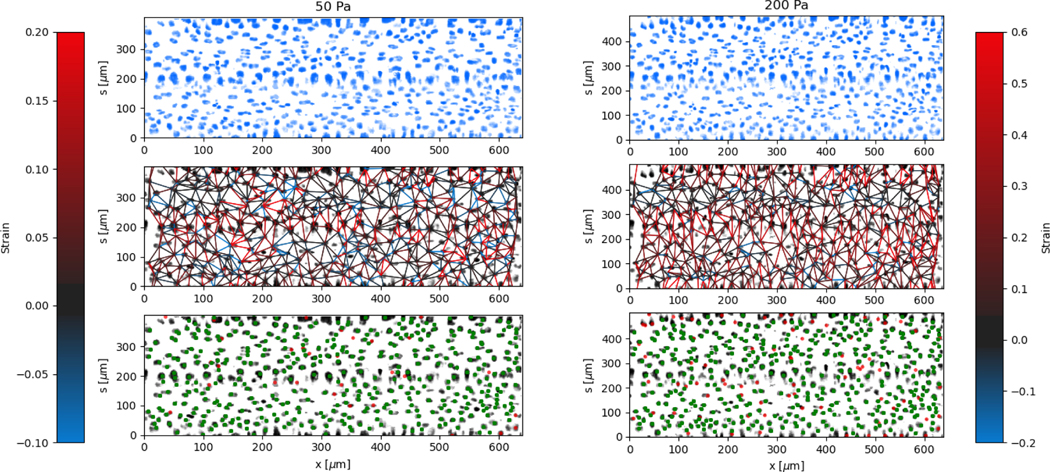
We use a three-dimensional (3D) microvascular platform to measure the elasticity and membrane permeability of the endothelial cell layer. The microfluidic platform is connected with a pneumatic pressure controller to apply hydrostatic pressure. The deformation is measured by tracking the mean vessel diameter under varying pressures up to 300 Pa. We obtain a value for the Young's modulus of the cell layer in low strain where a linear elastic response is observed and use a hyperelastic model that describes the strain hardening observed at larger strains (pressure). A fluorescent dye is used to track the flow through the cell layer to determine the membrane flow resistance as a function of applied pressure. Finally, we track the 3D positions of cell nuclei while the vessel is pressurized to observe local deformation and correlate inter-cell deformation with the local structure of the cell layer. This approach is able to probe the mechanical properties of blood vessels and provides a methodology for investigating microvascular related diseases.
我们使用三维(3D)微血管平台来测量内皮细胞层的弹性和膜通透性。微流控平台与气动压力控制器相连,以施加静水压力。通过在高达 300Pa 的不同压力下跟踪平均血管直径来测量变形。在观察到线性弹性响应的低应变下,我们获得细胞层的杨氏模量值,并使用描述在较大应变(压力)下观察到的应变硬化的超弹性模型。荧光染料用于跟踪流过细胞层的流动,以确定作为施加压力函数的膜流动阻力。最后,我们在血管加压时跟踪细胞核的 3D 位置,以观察局部变形,并将细胞间变形与细胞层的局部结构相关联。这种方法能够探测血管的机械性能,并为研究微血管相关疾病提供了一种方法。