• 文献检索
  • 文档翻译
  • 深度研究
  • 学术资讯
  • Suppr Zotero 插件Zotero 插件
  • 邀请有礼
  • 套餐&价格
  • 历史记录
应用&插件
Suppr Zotero 插件Zotero 插件浏览器插件Mac 客户端Windows 客户端微信小程序
定价
高级版会员购买积分包购买API积分包
服务
文献检索文档翻译深度研究API 文档MCP 服务
关于我们
关于 Suppr公司介绍联系我们用户协议隐私条款
关注我们

Suppr 超能文献

核心技术专利:CN118964589B侵权必究
粤ICP备2023148730 号-1Suppr @ 2026

文献检索

告别复杂PubMed语法,用中文像聊天一样搜索,搜遍4000万医学文献。AI智能推荐,让科研检索更轻松。

立即免费搜索

文件翻译

保留排版,准确专业,支持PDF/Word/PPT等文件格式,支持 12+语言互译。

免费翻译文档

深度研究

AI帮你快速写综述,25分钟生成高质量综述,智能提取关键信息,辅助科研写作。

立即免费体验

一种用于芯片上微血管形态分析和流速估计的三维方法。

A three-dimensional method for morphological analysis and flow velocity estimation in microvasculature on-a-chip.

作者信息

Rota Alberto, Possenti Luca, Offeddu Giovanni S, Senesi Martina, Stucchi Adelaide, Venturelli Irene, Rancati Tiziana, Zunino Paolo, Kamm Roger D, Costantino Maria Laura

机构信息

LaBS, Chemistry, Materials, and Chemical Engineering "Giulio Natta" Department Politecnico di Milano Milan Italy.

Data Science Unit, Department of Epidemiology and Data Science Fondazione IRCCS Istituto Nazionale dei Tumori Milan Italy.

出版信息

Bioeng Transl Med. 2023 Jun 11;8(5):e10557. doi: 10.1002/btm2.10557. eCollection 2023 Sep.

DOI:10.1002/btm2.10557
PMID:37693050
原文链接:https://pmc.ncbi.nlm.nih.gov/articles/PMC10487341/
Abstract

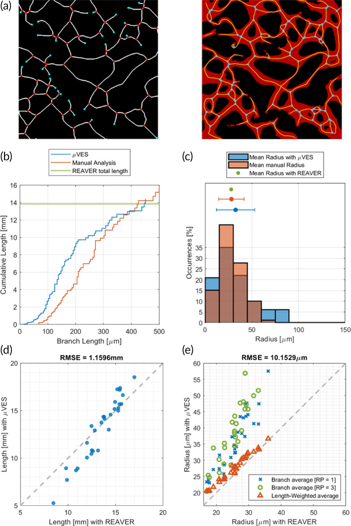
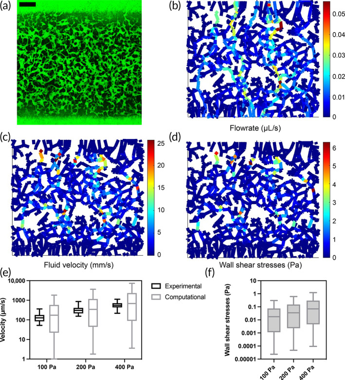
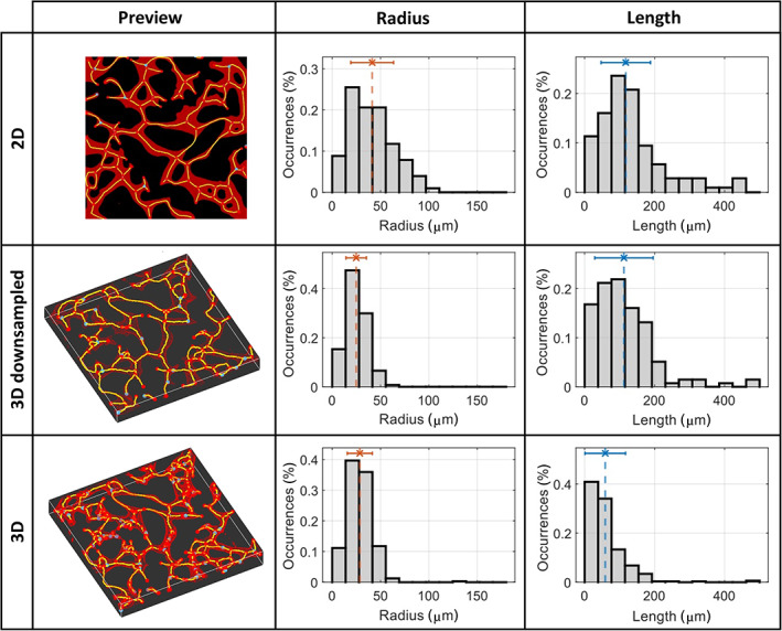
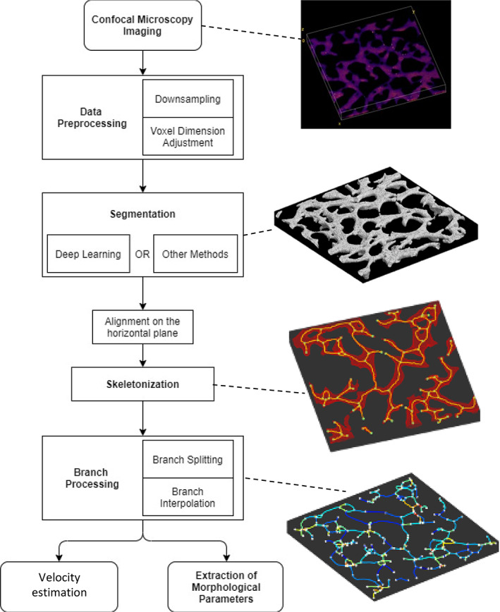
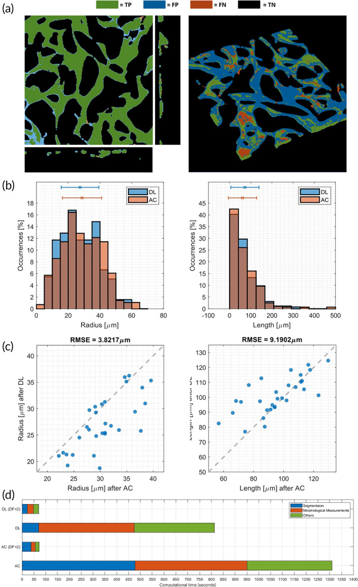
Three-dimensional (3D) imaging techniques (e.g., confocal microscopy) are commonly used to visualize in vitro models, especially microvasculature on-a-chip. Conversely, 3D analysis is not the standard method to extract quantitative information from those models. We developed the μVES algorithm to analyze vascularized in vitro models leveraging 3D data. It computes morphological parameters (geometry, diameter, length, tortuosity, eccentricity) and intravascular flow velocity. μVES application to microfluidic vascularized in vitro models shows that they successfully replicate functional features of the microvasculature in vivo in terms of intravascular fluid flow velocity. However, wall shear stress is lower compared to in vivo references. The morphological analysis also highlights the model's physiological similarities (vessel length and tortuosity) and shortcomings (vessel radius and surface-over-volume ratio). The addition of the third dimension in our analysis produced significant differences in the metrics assessed compared to 2D estimations. It enabled the computation of new indices, such as vessel eccentricity. These μVES capabilities can find application in analyses of different in vitro vascular models, as well as in vivo and ex vivo microvasculature.

摘要

三维(3D)成像技术(如共聚焦显微镜)通常用于可视化体外模型,尤其是芯片上的微血管系统。相反,3D分析并不是从这些模型中提取定量信息的标准方法。我们开发了μVES算法,利用3D数据来分析血管化体外模型。它可以计算形态学参数(几何形状、直径、长度、曲折度、偏心率)和血管内流速。将μVES应用于微流控血管化体外模型表明,就血管内流体流速而言,它们成功地复制了体内微血管系统的功能特征。然而,与体内参考值相比,壁面剪应力较低。形态学分析还突出了该模型的生理相似性(血管长度和曲折度)和不足之处(血管半径和表面积与体积比)。与二维估计相比,我们分析中增加的第三维在评估指标上产生了显著差异。它使得能够计算新的指标,如血管偏心率。这些μVES功能可应用于不同体外血管模型的分析,以及体内和离体微血管系统的分析。

https://cdn.ncbi.nlm.nih.gov/pmc/blobs/1e2a/10487341/6fde29a684d1/BTM2-8-e10557-g005.jpg
https://cdn.ncbi.nlm.nih.gov/pmc/blobs/1e2a/10487341/659d0564d84b/BTM2-8-e10557-g003.jpg
https://cdn.ncbi.nlm.nih.gov/pmc/blobs/1e2a/10487341/8bbbcaced1af/BTM2-8-e10557-g002.jpg
https://cdn.ncbi.nlm.nih.gov/pmc/blobs/1e2a/10487341/b3df03300582/BTM2-8-e10557-g001.jpg
https://cdn.ncbi.nlm.nih.gov/pmc/blobs/1e2a/10487341/c3d4286c88d8/BTM2-8-e10557-g004.jpg
https://cdn.ncbi.nlm.nih.gov/pmc/blobs/1e2a/10487341/6fde29a684d1/BTM2-8-e10557-g005.jpg

相似文献

1
A three-dimensional method for morphological analysis and flow velocity estimation in microvasculature on-a-chip.一种用于芯片上微血管形态分析和流速估计的三维方法。
Bioeng Transl Med. 2023 Jun 11;8(5):e10557. doi: 10.1002/btm2.10557. eCollection 2023 Sep.
2
Quantitative and Qualitative Comparison of 4D-DSA with 3D-DSA Using Computational Fluid Dynamics Simulations in Cerebral Aneurysms.基于计算流体动力学模拟的脑动脉瘤 4D-DSA 与 3D-DSA 的定量与定性比较。
AJNR Am J Neuroradiol. 2019 Sep;40(9):1505-1510. doi: 10.3174/ajnr.A6172.
3
Contrast-enhanced micro-CT imaging in murine carotid arteries: a new protocol for computing wall shear stress.小鼠颈动脉的对比增强微型CT成像:一种计算壁面剪应力的新方案
Biomed Eng Online. 2016 Dec 28;15(Suppl 2):156. doi: 10.1186/s12938-016-0270-2.
4
Mimicking arterial thrombosis in a 3D-printed microfluidic in vitro vascular model based on computed tomography angiography data.基于 CT 血管造影数据的 3D 打印微流控体外血管模型中模拟动脉血栓形成。
Lab Chip. 2017 Aug 8;17(16):2785-2792. doi: 10.1039/c7lc00202e.
5
Automatic segmentation, feature extraction and comparison of healthy and stroke cerebral vasculature.自动分割、特征提取和健康与中风脑血管的比较。
Neuroimage Clin. 2021;30:102573. doi: 10.1016/j.nicl.2021.102573. Epub 2021 Jan 26.
6
Quantification of Morphological Features in Non-Contrast-Enhanced Ultrasound Microvasculature Imaging.非增强超声微血管成像中形态学特征的量化
IEEE Access. 2020;8:18925-18937. doi: 10.1109/ACCESS.2020.2968292. Epub 2020 Jan 21.
7
Accurate noninvasive quantitation of blood flow, cross-sectional lumen vessel area and wall shear stress by three-dimensional paraboloid modeling of magnetic resonance imaging velocity data.通过磁共振成像速度数据的三维抛物面建模对血流、血管横截面积和壁面剪应力进行准确的无创定量分析。
J Am Coll Cardiol. 1998 Jul;32(1):128-34. doi: 10.1016/s0735-1097(98)00207-1.
8
Vascularized microfluidic platforms to mimic the tumor microenvironment.血管化微流控平台模拟肿瘤微环境。
Biotechnol Bioeng. 2018 Nov;115(11):2793-2806. doi: 10.1002/bit.26778. Epub 2018 Sep 6.
9
Capturing physiological hemodynamic flow and mechanosensitive cell signaling in vessel-on-a-chip platforms.在芯片上血管平台中捕获生理血液动力学流动和机械敏感细胞信号传导。
Front Physiol. 2024 Jul 29;15:1425618. doi: 10.3389/fphys.2024.1425618. eCollection 2024.
10
Reconstruction of blood propagation in three-dimensional rotational X-ray angiography (3D-RA).三维旋转X射线血管造影术(3D-RA)中血液传播的重建。
Comput Med Imaging Graph. 2005 Oct;29(7):507-20. doi: 10.1016/j.compmedimag.2005.03.006. Epub 2005 Sep 2.

引用本文的文献

1
Methods for processing and analyzing images of vascularized micro-organ and tumor systems.血管化微生物和肿瘤系统图像的处理与分析方法。
Front Bioeng Biotechnol. 2025 Jun 12;13:1585003. doi: 10.3389/fbioe.2025.1585003. eCollection 2025.
2
Remodeling of self-assembled microvascular networks under long term flow.长期流动作用下自组装微血管网络的重塑
bioRxiv. 2025 Mar 18:2025.03.17.643791. doi: 10.1101/2025.03.17.643791.
3
Methods for Processing and Analyzing Images of Vascularized Micro-Organ and Tumor Systems.血管化微器官和肿瘤系统图像的处理与分析方法

本文引用的文献

1
Aging-related cerebral microvascular changes visualized using ultrasound localization microscopy in the living mouse.利用活体小鼠超声定位显微镜观察与衰老相关的脑微血管变化。
Sci Rep. 2022 Jan 12;12(1):619. doi: 10.1038/s41598-021-04712-8.
2
Blood vessel-on-a-chip examines the biomechanics of microvasculature.血管芯片检测微血管的生物力学。
Soft Matter. 2021 Dec 22;18(1):117-125. doi: 10.1039/d1sm01312b.
3
3D Biofabricated In Vitro Models of Vascularized and Mineralized Bone Tissues.血管化和矿化骨组织的3D生物制造体外模型
bioRxiv. 2025 Mar 14:2025.03.12.642741. doi: 10.1101/2025.03.12.642741.
4
Three-dimensional modeling of flow through microvascular beds and surrounding interstitial spaces.通过微血管床和周围间质空间的血流三维建模。
bioRxiv. 2025 Feb 15:2024.02.28.582152. doi: 10.1101/2024.02.28.582152.
5
Vascularized organoid-on-a-chip: design, imaging, and analysis.血管化类器官芯片:设计、成像和分析。
Angiogenesis. 2024 May;27(2):147-172. doi: 10.1007/s10456-024-09905-z. Epub 2024 Feb 26.
Methods Mol Biol. 2022;2373:283-296. doi: 10.1007/978-1-0716-1693-2_17.
4
Engineering new microvascular networks on-chip: ingredients, assembly, and best practices.在芯片上构建新型微血管网络:要素、组装及最佳实践
Adv Funct Mater. 2021 Apr 1;31(14). doi: 10.1002/adfm.202007199. Epub 2021 Jan 20.
5
3D Imaging and Quantitative Characterization of Mouse Capillary Coronary Network Architecture.小鼠冠状动脉毛细血管网络结构的三维成像与定量表征
Biology (Basel). 2021 Apr 7;10(4):306. doi: 10.3390/biology10040306.
6
Review on the Vascularization of Organoids and Organoids-on-a-hip.类器官及类器官芯片血管化研究综述
Front Bioeng Biotechnol. 2021 Apr 12;9:637048. doi: 10.3389/fbioe.2021.637048. eCollection 2021.
7
Vascularized Microfluidics and Their Untapped Potential for Discovery in Diseases of the Microvasculature.血管化微流控及其在微血管疾病发现中的未开发潜力。
Annu Rev Biomed Eng. 2021 Jul 13;23:407-432. doi: 10.1146/annurev-bioeng-091520-025358. Epub 2021 Apr 16.
8
Engineering Vascularized Organoid-on-a-Chip Models.工程化血管化类器官芯片模型。
Annu Rev Biomed Eng. 2021 Jul 13;23:141-167. doi: 10.1146/annurev-bioeng-090120-094330. Epub 2021 Mar 23.
9
Engineering the early bone metastatic niche through human vascularized immuno bone minitissues.通过人源化血管化免疫骨微组织工程构建早期骨转移龛。
Biofabrication. 2021 Apr 26;13(3). doi: 10.1088/1758-5090/abefea.
10
The cancer glycocalyx mediates intravascular adhesion and extravasation during metastatic dissemination.肿瘤糖萼在转移扩散过程中介导血管内黏附和血管外渗。
Commun Biol. 2021 Feb 26;4(1):255. doi: 10.1038/s42003-021-01774-2.