Zhang Xiao, Liu Yang, Zuo Qiang, Wang Qingyun, Li Zuxi, Yan Kai, Yuan Tao, Zhang Yi, Shen Kai, Xie Rui, Fan Weimin
Department of Orthopedics, The First Affiliated Hospital of Nanjing Medical University, Nanjing, China.
Int J Bioprint. 2021 Sep 14;7(4):401. doi: 10.18063/ijb.v7i4.401. eCollection 2021.
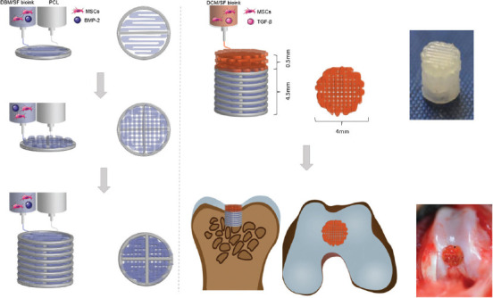
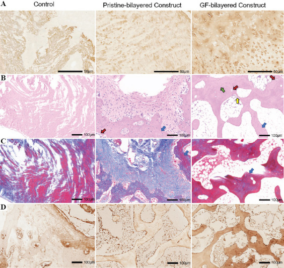
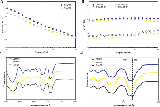
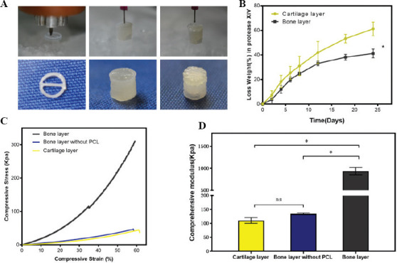
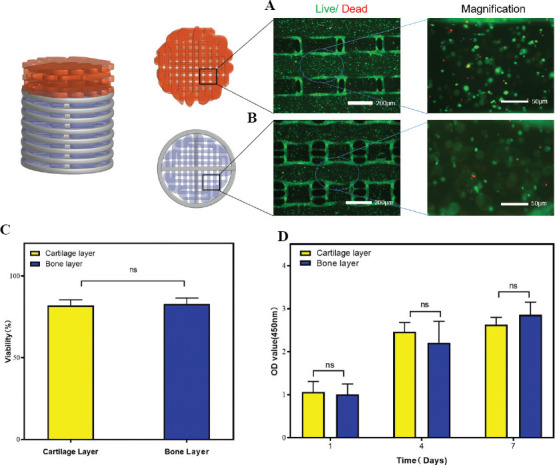
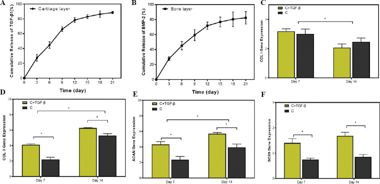
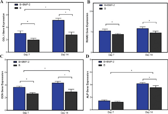
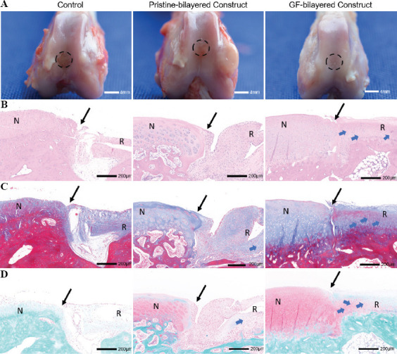
Recently, three-dimensional (3D) bioprinting technology is becoming an appealing approach for osteochondral repair. However, it is challenging to develop a bilayered scaffold with anisotropic structural properties to mimic a native osteochondral tissue. Herein, we developed a bioink consisting of decellularized extracellular matrix and silk fibroin to print the bilayered scaffold. The bilayered scaffold mimics the natural osteochondral tissue by controlling the composition, mechanical properties, and growth factor release in each layer of the scaffold. The results show that each layer of scaffolds had a suitable mechanical strength and degradation rate. Furthermore, the scaffolds encapsulating transforming growth factor-beta (TGF-β) and bone morphogenetic protein-2 (BMP-2) can act as a controlled release system and promote directed differentiation of bone marrow-derived mesenchymal stem cells. Furthermore, the experiments suggested that the scaffolds loaded with growth factors promoted osteochondral regeneration in the rabbit knee joint model. Consequently, the biomimetic bilayered scaffold loaded with TGF-β and BMP-2 would be a promising strategy for osteochondral repair.
最近,三维(3D)生物打印技术正成为一种用于骨软骨修复的有吸引力的方法。然而,开发一种具有各向异性结构特性的双层支架以模拟天然骨软骨组织具有挑战性。在此,我们开发了一种由脱细胞细胞外基质和丝素蛋白组成的生物墨水来打印双层支架。该双层支架通过控制支架各层的组成、力学性能和生长因子释放来模拟天然骨软骨组织。结果表明,支架的每一层都具有合适的机械强度和降解速率。此外,封装转化生长因子-β(TGF-β)和骨形态发生蛋白-2(BMP-2)的支架可作为控释系统,并促进骨髓间充质干细胞的定向分化。此外,实验表明,负载生长因子的支架在兔膝关节模型中促进了骨软骨再生。因此,负载TGF-β和BMP-2的仿生双层支架将是一种有前景的骨软骨修复策略。