Division of Electronic Systems and Signal Processing, Institute of Automatic Control and Robotics, Poznan University of Technology, 60-965 Poznan, Poland.
Department of Ophthalmology, Chair of Ophthalmology and Optometry, Heliodor Swiecicki University Hospital, Poznan University of Medical Sciences, 60-780 Poznan, Poland.
Sensors (Basel). 2021 Nov 12;21(22):7521. doi: 10.3390/s21227521.
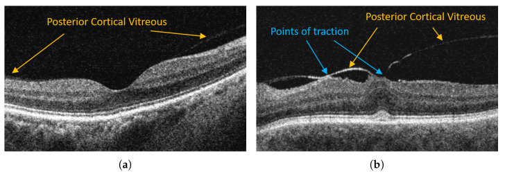
This paper proposes an efficient segmentation of the preretinal area between the inner limiting membrane (ILM) and posterior cortical vitreous (PCV) of the human eye in an image obtained with the use of optical coherence tomography (OCT). The research was carried out using a database of three-dimensional OCT imaging scans obtained with the Optovue RTVue XR Avanti device. Various types of neural networks (UNet, Attention UNet, ReLayNet, LFUNet) were tested for semantic segmentation, their effectiveness was assessed using the Dice coefficient and compared to the graph theory techniques. Improvement in segmentation efficiency was achieved through the use of relative distance maps. We also show that selecting a larger kernel size for convolutional layers can improve segmentation quality depending on the neural network model. In the case of PVC, we obtain the effectiveness reaching up to 96.35%. The proposed solution can be widely used to diagnose vitreomacular traction changes, which is not yet available in scientific or commercial OCT imaging solutions.
本文提出了一种在使用光学相干断层扫描(OCT)获得的图像中对视网膜前区域(ILM 和 PCV 之间)进行有效分割的方法。该研究使用 Optovue RTVue XR Avanti 设备获得的三维 OCT 成像扫描数据库进行。针对语义分割测试了各种类型的神经网络(UNet、Attention UNet、ReLayNet、LFUNet),使用 Dice 系数评估了它们的有效性,并与图论技术进行了比较。通过使用相对距离图,可以提高分割效率。我们还表明,根据神经网络模型,选择更大的卷积层核大小可以提高分割质量。在 PVC 的情况下,我们获得了高达 96.35%的有效性。所提出的解决方案可以广泛用于诊断玻璃体黄斑牵引变化,而目前科学或商业 OCT 成像解决方案还无法实现这一点。