Center for Neurotechnology and Neurorecovery, Department of Neurology, Massachusetts General Hospital and Harvard Medical School, Boston, MA, USA; Athinoula A. Martinos Center for Biomedical Imaging, Massachusetts General Hospital and Harvard Medical School, Charlestown, MA, USA.
Center for Neurotechnology and Neurorecovery, Department of Neurology, Massachusetts General Hospital and Harvard Medical School, Boston, MA, USA; Harvard Medical School, Boston, MA, USA.
Neuroimage. 2021 Dec 15;245:118758. doi: 10.1016/j.neuroimage.2021.118758. Epub 2021 Nov 25.
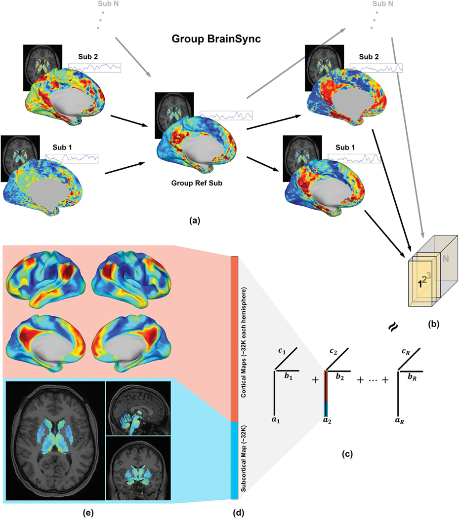
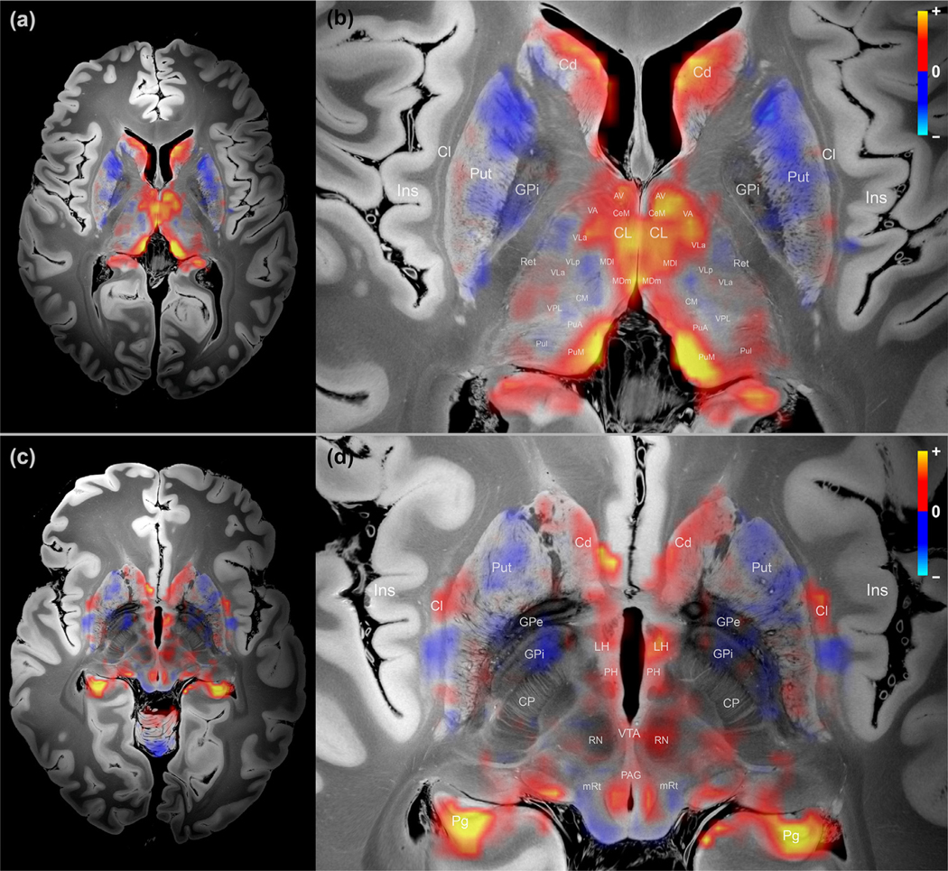
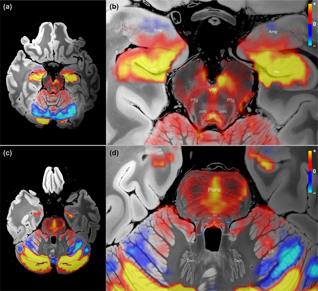
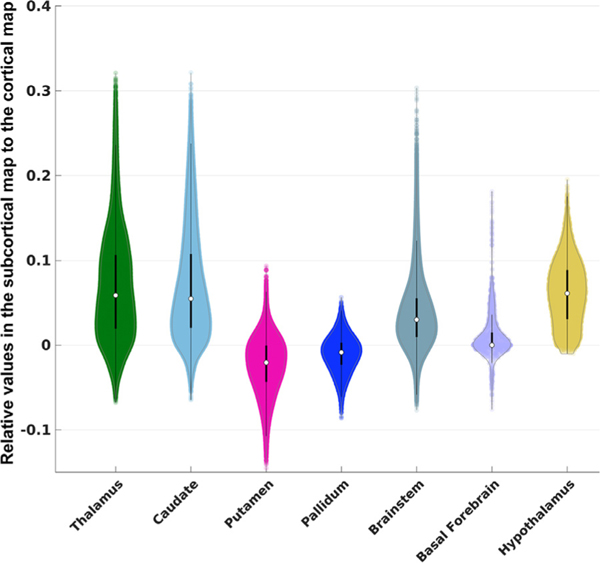
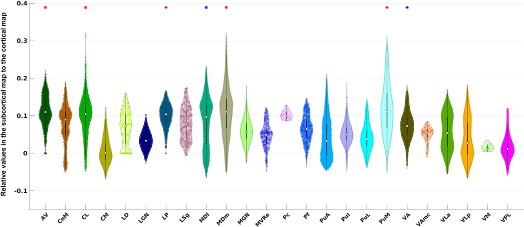
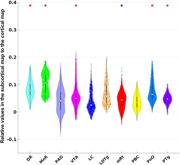
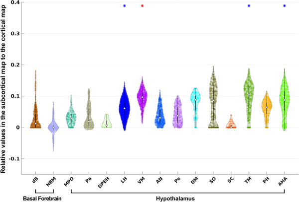
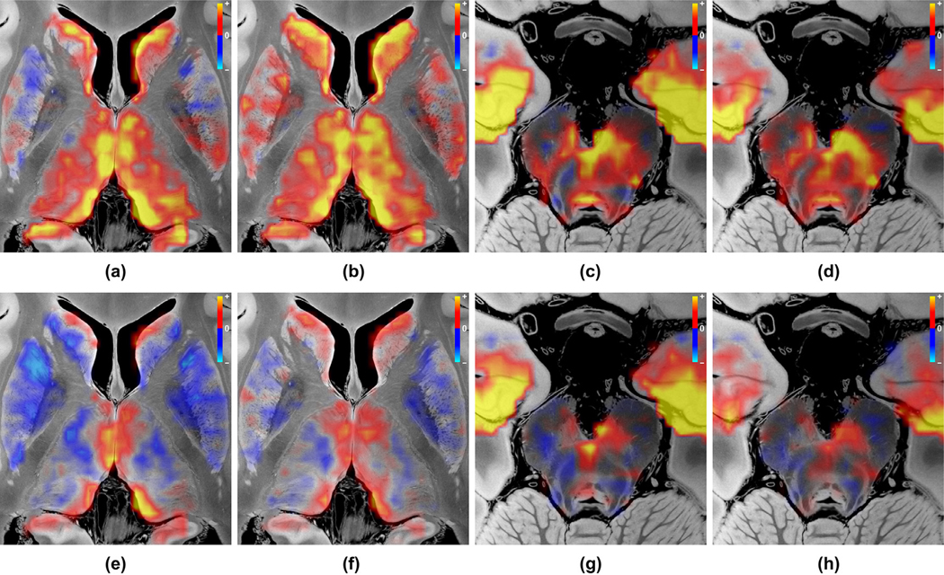
The default mode network (DMN) mediates self-awareness and introspection, core components of human consciousness. Therapies to restore consciousness in patients with severe brain injuries have historically targeted subcortical sites in the brainstem, thalamus, hypothalamus, basal forebrain, and basal ganglia, with the goal of reactivating cortical DMN nodes. However, the subcortical connectivity of the DMN has not been fully mapped, and optimal subcortical targets for therapeutic neuromodulation of consciousness have not been identified. In this work, we created a comprehensive map of DMN subcortical connectivity by combining high-resolution functional and structural datasets with advanced signal processing methods. We analyzed 7 Tesla resting-state functional MRI (rs-fMRI) data from 168 healthy volunteers acquired in the Human Connectome Project. The rs-fMRI blood-oxygen-level-dependent (BOLD) data were temporally synchronized across subjects using the BrainSync algorithm. Cortical and subcortical DMN nodes were jointly analyzed and identified at the group level by applying a novel Nadam-Accelerated SCAlable and Robust (NASCAR) tensor decomposition method to the synchronized dataset. The subcortical connectivity map was then overlaid on a 7 Tesla 100 µm ex vivo MRI dataset for neuroanatomic analysis using automated segmentation of nuclei within the brainstem, thalamus, hypothalamus, basal forebrain, and basal ganglia. We further compared the NASCAR subcortical connectivity map with its counterpart generated from canonical seed-based correlation analyses. The NASCAR method revealed that BOLD signal in the central lateral nucleus of the thalamus and ventral tegmental area of the midbrain is strongly correlated with that of the DMN. In an exploratory analysis, additional subcortical sites in the median and dorsal raphe, lateral hypothalamus, and caudate nuclei were correlated with the cortical DMN. We also found that the putamen and globus pallidus are negatively correlated (i.e., anti-correlated) with the DMN, providing rs-fMRI evidence for the mesocircuit hypothesis of human consciousness, whereby a striatopallidal feedback system modulates anterior forebrain function via disinhibition of the central thalamus. Seed-based analyses yielded similar subcortical DMN connectivity, but the NASCAR result showed stronger contrast and better spatial alignment with dopamine immunostaining data. The DMN subcortical connectivity map identified here advances understanding of the subcortical regions that contribute to human consciousness and can be used to inform the selection of therapeutic targets in clinical trials for patients with disorders of consciousness.
默认模式网络 (DMN) 介导自我意识和内省,这是人类意识的核心组成部分。历史上,用于恢复严重脑损伤患者意识的疗法靶向于脑干、丘脑、下丘脑、基底前脑和基底神经节中的皮质下部位,目的是重新激活皮质 DMN 节点。然而,DMN 的皮质下连接尚未完全绘制,也没有确定用于意识治疗性神经调节的最佳皮质下靶标。在这项工作中,我们通过结合高分辨率功能和结构数据集以及先进的信号处理方法,创建了 DMN 皮质下连接的综合图谱。我们分析了来自人类连接组计划的 168 名健康志愿者的 7 特斯拉静息状态功能磁共振成像 (rs-fMRI) 数据。使用 BrainSync 算法对 rs-fMRI 血氧水平依赖 (BOLD) 数据进行了跨受试者的时间同步。通过应用一种新颖的基于 Nesterov 加速的可扩展和鲁棒 (NASCAR) 张量分解方法对同步数据集进行分析,在组水平上共同分析和确定皮质和皮质下 DMN 节点。然后,将皮质下连接图叠加在 7 特斯拉 100 µm 离体 MRI 数据集上,使用脑干部、丘脑、下丘脑、基底前脑和基底神经节内核的自动分割进行神经解剖学分析。我们还比较了 NASCAR 皮质下连接图与其基于经典种子相关分析生成的对应图。NASCAR 方法表明,丘脑中央外侧核和中脑腹侧被盖区的 BOLD 信号与 DMN 强烈相关。在探索性分析中,中缝核和背侧中缝核、外侧下丘脑和尾状核中的其他皮质下部位与皮质 DMN 相关。我们还发现纹状体和苍白球与 DMN 呈负相关(即,反相关),为人类意识的中脑回路假说提供了 rs-fMRI 证据,其中纹状体-苍白球反馈系统通过抑制中央丘脑来调节前脑功能。种子分析得出了相似的皮质下 DMN 连接,但 NASCAR 结果显示与多巴胺免疫染色数据的对比度更强,空间对齐更好。这里确定的 DMN 皮质下连接图谱推进了对有助于人类意识的皮质下区域的理解,并可用于为意识障碍患者的临床试验中治疗靶标的选择提供信息。