Yan Li, Bai Chen, Zheng Yu, Zhou Xiaodong, Wan Mingxi, Zong Yujin, Chen Shanshan, Zhou Yin
Institute of Medical Research, Northwestern Polytechnical University, Xi'an, China.
State Key Laboratory of Transient Optics and Photonics, Xi'an Institute of Optics and Precision Mechanics, Chinese Academy of Sciences, Xi'an, China.
Front Neurol. 2021 Nov 17;12:720320. doi: 10.3389/fneur.2021.720320. eCollection 2021.
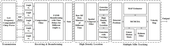
Ultrasound is ideal for displaying intracranial great vessels but not intracranial microvessels and terminal vessels. Even with contrast agents, the imaging effect is still unsatisfactory. In recent years, significant theoretical advances have been achieved in super-resolution imaging. The latest commonly used ultrafast plane-wave ultrasound Doppler imaging of the brain and microbubble-based super-resolution ultrasound imaging have been applied to the imaging of cerebral microvessels and blood flow in small animals such as mice but have not been applied to imaging of the cerebral microvessels in monkeys and larger animals. In China, preliminary research results have been obtained using super-resolution imaging in certain fields but rarely in fundamental and clinical experiments on large animals. In recent years, we have conducted a joint study with the Xi'an Jiaotong University to explore the application and performance of this new technique in the diagnosis of cerebrovascular diseases in large animals. To explore the characteristics and advantages of microbubble-based super-resolution ultrasound imaging of intracranial vessels in rhesus monkeys compared with conventional transcranial ultrasound. First, the effectiveness and feasibility of the super-resolution imaging technique were verified by modular simulation experiments. Then, the imaging parameters were adjusted based on experiments. Finally, two rhesus monkeys were used for experiments of intracranial microvessel imaging. Compared with conventional plane-wave imaging, super-resolution imaging could measure the inner diameters of cerebral microvessels at a resolution of 1 mm or even 0.7 mm and extract blood flow information. In addition, it has a better signal-to-noise ratio (5.625 dB higher) and higher resolution (~30-fold higher). The results of the experiments with rhesus monkeys showed that microbubble-based super-resolution ultrasound imaging can achieve an optimal resolution at the micron level and an imaging depth >35 mm. Super-resolution imaging can realize the monitoring imaging of high-resolution and fast calculation of microbubbles in the process of tissue damage, providing an important experimental basis for the clinical application of non-invasive transcranial ultrasound.
超声对于显示颅内大血管是理想的,但对于颅内微血管和终末血管则不然。即使使用造影剂,成像效果仍然不尽人意。近年来,超分辨率成像取得了重大理论进展。最新常用的脑超快平面波超声多普勒成像和基于微泡的超分辨率超声成像已应用于小鼠等小动物的脑微血管成像和血流成像,但尚未应用于猴子和大型动物的脑微血管成像。在中国,在某些领域使用超分辨率成像已取得初步研究成果,但在大型动物的基础和临床实验中却很少见。近年来,我们与西安交通大学开展了联合研究,以探索这项新技术在大型动物脑血管疾病诊断中的应用和性能。为了探讨与传统经颅超声相比,基于微泡的恒河猴颅内血管超分辨率超声成像的特点和优势。首先,通过模块模拟实验验证了超分辨率成像技术的有效性和可行性。然后,根据实验调整成像参数。最后,使用两只恒河猴进行颅内微血管成像实验。与传统平面波成像相比,超分辨率成像能够以1毫米甚至0.7毫米的分辨率测量脑微血管内径并提取血流信息。此外,它具有更好的信噪比(高5.625分贝)和更高的分辨率(高约30倍)。恒河猴的实验结果表明,基于微泡的超分辨率超声成像可以在微米水平实现最佳分辨率,成像深度>35毫米。超分辨率成像能够在组织损伤过程中实现对微泡的高分辨率和快速计算的监测成像,为无创经颅超声的临床应用提供重要的实验依据。