• 文献检索
  • 文档翻译
  • 深度研究
  • 学术资讯
  • Suppr Zotero 插件Zotero 插件
  • 邀请有礼
  • 套餐&价格
  • 历史记录
应用&插件
Suppr Zotero 插件Zotero 插件浏览器插件Mac 客户端Windows 客户端微信小程序
定价
高级版会员购买积分包购买API积分包
服务
文献检索文档翻译深度研究API 文档MCP 服务
关于我们
关于 Suppr公司介绍联系我们用户协议隐私条款
关注我们

Suppr 超能文献

核心技术专利:CN118964589B侵权必究
粤ICP备2023148730 号-1Suppr @ 2026

文献检索

告别复杂PubMed语法,用中文像聊天一样搜索,搜遍4000万医学文献。AI智能推荐,让科研检索更轻松。

立即免费搜索

文件翻译

保留排版,准确专业,支持PDF/Word/PPT等文件格式,支持 12+语言互译。

免费翻译文档

深度研究

AI帮你快速写综述,25分钟生成高质量综述,智能提取关键信息,辅助科研写作。

立即免费体验

脂肪抑制T2加权磁共振成像的纹理分析及利用机器学习鉴别鼻腔和鼻窦小圆形恶性细胞肿瘤

Texture Analysis of Fat-Suppressed T2-Weighted Magnetic Resonance Imaging and Use of Machine Learning to Discriminate Nasal and Paranasal Sinus Small Round Malignant Cell Tumors.

作者信息

Chen Chen, Qin Yuhui, Cheng Junying, Gao Fabao, Zhou Xiaoyue

机构信息

Department of Radiology, West China Hospital, Sichuan University, Chengdu, China.

Department of MRI, The First Affiliated Hospital of Zhengzhou University, Zhengzhou, China.

出版信息

Front Oncol. 2021 Dec 13;11:701289. doi: 10.3389/fonc.2021.701289. eCollection 2021.

DOI:10.3389/fonc.2021.701289
PMID:34966664
原文链接:https://pmc.ncbi.nlm.nih.gov/articles/PMC8710453/
Abstract

OBJECTIVE

We used texture analysis and machine learning (ML) to classify small round cell malignant tumors (SRCMTs) and Non-SRCMTs of nasal and paranasal sinus on fat-suppressed T2 weighted imaging (Fs-T2WI).

MATERIALS

Preoperative MRI scans of 164 patients from 1 January 2018 to 1 January 2021 diagnosed with SRCMTs and Non-SRCMTs were included in this study. A total of 271 features were extracted from each regions of interest. Datasets were randomly divided into two sets, including a training set (∼70%) and a test set (∼30%). The Pearson correlation coefficient (PCC) and principal component analysis (PCA) methods were performed to reduce dimensions, and the Analysis of Variance (ANOVA), Kruskal-Wallis (KW), and Recursive Feature Elimination (RFE) and Relief were performed for feature selections. Classifications were performed using 10 ML classifiers. Results were evaluated using a leave one out cross-validation analysis.

RESULTS

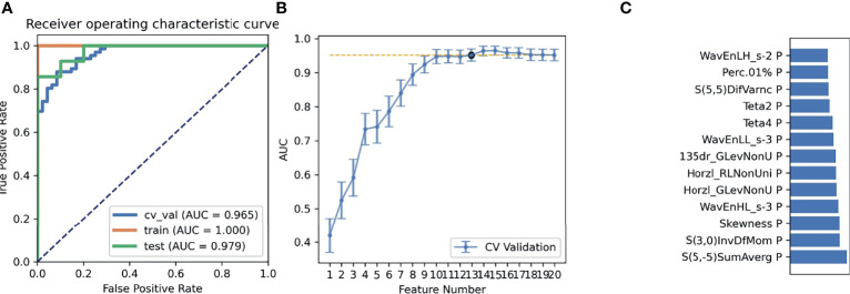
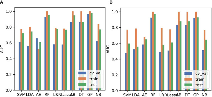
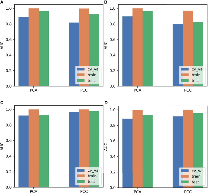
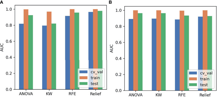
We compared the AUC of all pipelines on the validation dataset with FeAture Explorer (FAE) software. The pipeline using a PCC dimension reduction, relief feature selection, and gaussian process (GP) classifier yielded the highest area under the curve (AUC) using 15 features. When the "one-standard error" rule was used, FAE also produced a simpler model with 13 features, including S(5,-5)SumAverg, S(3,0)InvDfMom, Skewness, WavEnHL_s-3, Horzl_GlevNonU, Horzl_RLNonUni, 135dr_GlevNonU, WavEnLL_s-3, Teta4, Teta2, S(5,5)DifVarnc, Perc.01%, and WavEnLH_s-2. The AUCs of the training/validation/test datasets were 1.000/0.965/0.979, and the accuracies, sensitivities, and specificities were 0.890, 0.880, and 0.920, respectively. The best algorithm was GP whose AUCs of the training/validation/test datasets by the two-dimensional reduction methods and four feature selection methods were greater than approximately 0.800. Especially, the AUCs of different datasets were greater than approximately 0.900 using the PCC, RFE/Relief, and GP algorithms.

CONCLUSIONS

We demonstrated the feasibility of combining artificial intelligence and the radiomics from Fs-T2WI to differentially diagnose SRCMTs and Non-SRCMTs. This non-invasive approach could be very promising in clinical oncology.

摘要

目的

我们运用纹理分析和机器学习(ML)对鼻腔及鼻窦的小圆形细胞恶性肿瘤(SRCMT)和非SRCMT在脂肪抑制T2加权成像(Fs-T2WI)上进行分类。

材料

本研究纳入了2018年1月1日至2021年1月1日期间164例被诊断为SRCMT和非SRCMT患者的术前MRI扫描图像。从每个感兴趣区域提取了总共271个特征。数据集被随机分为两组,包括训练集(约70%)和测试集(约30%)。采用Pearson相关系数(PCC)和主成分分析(PCA)方法进行降维,并进行方差分析(ANOVA)、Kruskal-Wallis(KW)、递归特征消除(RFE)和Relief进行特征选择。使用10种ML分类器进行分类。结果采用留一法交叉验证分析进行评估。

结果

我们使用FeAture Explorer(FAE)软件在验证数据集上比较了所有流程的曲线下面积(AUC)。使用PCC降维、Relief特征选择和高斯过程(GP)分类器的流程在使用15个特征时产生了最高的曲线下面积(AUC)。当使用“一个标准误差”规则时,FAE还生成了一个更简单的包含13个特征的模型,包括S(5,-5)SumAverg、S(3,0)InvDfMom、偏度、WavEnHL_s-3、Horzl_GlevNonU、Horzl_RLNonUni、135dr_GlevNonU、WavEnLL_s-3、Teta4、Teta2、S(5,5)DifVarnc、Perc.01%和WavEnLH_s-2。训练/验证/测试数据集的AUC分别为1.000/0.965/0.979,准确率、敏感性和特异性分别为0.890、0.880和0.920。最佳算法是GP,其通过二维降维方法和四种特征选择方法在训练/验证/测试数据集上的AUC大于约0.800。特别是,使用PCC、RFE/Relief和GP算法时不同数据集的AUC大于约0.900。

结论

我们证明了结合人工智能和Fs-T2WI的放射组学对SRCMT和非SRCMT进行鉴别诊断的可行性。这种非侵入性方法在临床肿瘤学中可能非常有前景。

https://cdn.ncbi.nlm.nih.gov/pmc/blobs/a019/8710453/4fd3d312e39f/fonc-11-701289-g004.jpg
https://cdn.ncbi.nlm.nih.gov/pmc/blobs/a019/8710453/8a516b1dac18/fonc-11-701289-g001.jpg
https://cdn.ncbi.nlm.nih.gov/pmc/blobs/a019/8710453/59e18f083faa/fonc-11-701289-g002.jpg
https://cdn.ncbi.nlm.nih.gov/pmc/blobs/a019/8710453/d585d701cc83/fonc-11-701289-g003.jpg
https://cdn.ncbi.nlm.nih.gov/pmc/blobs/a019/8710453/4fd3d312e39f/fonc-11-701289-g004.jpg

相似文献

1
Texture Analysis of Fat-Suppressed T2-Weighted Magnetic Resonance Imaging and Use of Machine Learning to Discriminate Nasal and Paranasal Sinus Small Round Malignant Cell Tumors.脂肪抑制T2加权磁共振成像的纹理分析及利用机器学习鉴别鼻腔和鼻窦小圆形恶性细胞肿瘤
Front Oncol. 2021 Dec 13;11:701289. doi: 10.3389/fonc.2021.701289. eCollection 2021.
2
Machine learning to differentiate small round cell malignant tumors and non-small round cell malignant tumors of the nasal and paranasal sinuses using apparent diffusion coefficient values.利用表观扩散系数值,通过机器学习鉴别鼻窦的小圆形细胞恶性肿瘤和非小圆形细胞恶性肿瘤。
Eur Radiol. 2022 Jun;32(6):3819-3829. doi: 10.1007/s00330-021-08465-w. Epub 2022 Jan 14.
3
Diagnostic Performance of 2D and 3D T2WI-Based Radiomics Features With Machine Learning Algorithms to Distinguish Solid Solitary Pulmonary Lesion.基于二维和三维T2加权成像的影像组学特征联合机器学习算法鉴别实性孤立性肺结节的诊断效能
Front Oncol. 2021 Nov 18;11:683587. doi: 10.3389/fonc.2021.683587. eCollection 2021.
4
Development and validation of a prediction model for malignant sinonasal tumors based on MR radiomics and machine learning.基于磁共振成像放射组学和机器学习的鼻窦恶性肿瘤预测模型的开发与验证
Eur Radiol. 2025 Apr;35(4):2074-2083. doi: 10.1007/s00330-024-11033-7. Epub 2024 Aug 30.
5
Classification of pulmonary lesion based on multiparametric MRI: utility of radiomics and comparison of machine learning methods.基于多参数 MRI 的肺部病变分类:放射组学的效用及机器学习方法的比较。
Eur Radiol. 2020 Aug;30(8):4595-4605. doi: 10.1007/s00330-020-06768-y. Epub 2020 Mar 28.
6
MRI-Based Grading of Clear Cell Renal Cell Carcinoma Using a Machine Learning Classifier.使用机器学习分类器基于MRI对透明细胞肾细胞癌进行分级
Front Oncol. 2021 Oct 1;11:708655. doi: 10.3389/fonc.2021.708655. eCollection 2021.
7
Bi-parametric magnetic resonance imaging based radiomics for the identification of benign and malignant prostate lesions: cross-vendor validation.基于双参数磁共振成像的放射组学用于鉴别前列腺良恶性病变:跨供应商验证。
Phys Eng Sci Med. 2021 Sep;44(3):745-754. doi: 10.1007/s13246-021-01022-1. Epub 2021 Jun 1.
8
The value of machine learning based on CT radiomics in the preoperative identification of peripheral nerve invasion in colorectal cancer: a two-center study.基于CT影像组学的机器学习在结直肠癌术前周围神经侵犯识别中的价值:一项双中心研究
Insights Imaging. 2024 Apr 5;15(1):101. doi: 10.1186/s13244-024-01664-1.
9
Multi-Parametric MRI-Based Radiomics Models for Predicting Molecular Subtype and Androgen Receptor Expression in Breast Cancer.基于多参数磁共振成像的放射组学模型预测乳腺癌分子亚型及雄激素受体表达
Front Oncol. 2021 Aug 18;11:706733. doi: 10.3389/fonc.2021.706733. eCollection 2021.
10
Intravoxel incoherent motion MRI for differentiating sinonasal small round cell malignant tumours (SRCMTs) from Non-SRCMTs: comparison and correlation with dynamic contrast-enhanced MRI.体素内不相干运动磁共振成像用于鉴别鼻窦小圆形细胞恶性肿瘤(SRCMTs)与非SRCMTs:与动态对比增强磁共振成像的比较及相关性
Clin Radiol. 2018 Nov;73(11):966-974. doi: 10.1016/j.crad.2018.07.097. Epub 2018 Aug 4.

引用本文的文献

1
Artificial intelligence and MRI in sinonasal tumors discrimination: where do we stand?人工智能与磁共振成像在鼻窦肿瘤鉴别中的应用:我们目前的进展如何?
Eur Arch Otorhinolaryngol. 2025 Mar;282(3):1557-1566. doi: 10.1007/s00405-024-09169-9. Epub 2024 Dec 24.
2
Classification of nasal polyps and inverted papillomas using CT-based radiomics.基于CT的放射组学对鼻息肉和内翻性乳头状瘤的分类
Insights Imaging. 2023 Nov 13;14(1):188. doi: 10.1186/s13244-023-01536-0.
3
Radiomics analysis based on CT for the prediction of pulmonary metastases in ewing sarcoma.

本文引用的文献

1
FeAture Explorer (FAE): A tool for developing and comparing radiomics models.特征探索器(FAE):一种用于开发和比较放射组学模型的工具。
PLoS One. 2020 Aug 17;15(8):e0237587. doi: 10.1371/journal.pone.0237587. eCollection 2020.
2
Positron emission computed tomography and magnetic resonance imaging features of sinonasal small round blue cell tumors.鼻窦小圆形蓝细胞肿瘤的正电子发射计算机断层扫描和磁共振成像特征
Neuroradiol J. 2020 Feb;33(1):48-56. doi: 10.1177/1971400919873895. Epub 2019 Aug 28.
3
Integration of morphological preprocessing and fractal based feature extraction with recursive feature elimination for skin lesion types classification.
基于 CT 的影像组学分析预测尤文肉瘤肺转移。
BMC Med Imaging. 2023 Oct 2;23(1):147. doi: 10.1186/s12880-023-01077-4.
4
Deep learning-enhanced radiomics for histologic classification and grade stratification of stage IA lung adenocarcinoma: a multicenter study.深度学习增强的放射组学用于IA期肺腺癌的组织学分类和分级分层:一项多中心研究
Front Oncol. 2023 Jul 20;13:1224455. doi: 10.3389/fonc.2023.1224455. eCollection 2023.
5
A Scoping Review of Artificial Intelligence Research in Rhinology.鼻科学人工智能研究的范围综述。
Am J Rhinol Allergy. 2023 Jul;37(4):438-448. doi: 10.1177/19458924231162437. Epub 2023 Mar 9.
6
Collinearity and Dimensionality Reduction in Radiomics: Effect of Preprocessing Parameters in Hypertrophic Cardiomyopathy Magnetic Resonance T1 and T2 Mapping.放射组学中的共线性和降维:肥厚型心肌病磁共振T1和T2映射中预处理参数的影响
Bioengineering (Basel). 2023 Jan 6;10(1):80. doi: 10.3390/bioengineering10010080.
7
Predicting IDH Mutation Status in Low-Grade Gliomas Based on Optimal Radiomic Features Combined with Multi-Sequence Magnetic Resonance Imaging.基于最优影像组学特征联合多序列磁共振成像预测低级别胶质瘤的异柠檬酸脱氢酶(IDH)突变状态
Diagnostics (Basel). 2022 Nov 30;12(12):2995. doi: 10.3390/diagnostics12122995.
8
Differentiating between cardiac amyloidosis and hypertrophic cardiomyopathy on non-contrast cine-magnetic resonance images using machine learning-based radiomics.使用基于机器学习的放射组学在非对比电影磁共振图像上鉴别心脏淀粉样变性和肥厚型心肌病。
Front Cardiovasc Med. 2022 Oct 26;9:1001269. doi: 10.3389/fcvm.2022.1001269. eCollection 2022.
9
Combination of pre-treatment dynamic [F]FET PET radiomics and conventional clinical parameters for the survival stratification in patients with IDH-wildtype glioblastoma.治疗前动态 [F]FET PET 影像组学与常规临床参数联合用于 IDH 野生型胶质母细胞瘤患者的生存分层。
Eur J Nucl Med Mol Imaging. 2023 Jan;50(2):535-545. doi: 10.1007/s00259-022-05988-2. Epub 2022 Oct 13.
形态学预处理与分形特征提取的集成以及递归特征消除在皮肤病变类型分类中的应用。
Comput Methods Programs Biomed. 2019 Sep;178:201-218. doi: 10.1016/j.cmpb.2019.06.018. Epub 2019 Jun 16.
4
Statistical characterization and classification of colon microarray gene expression data using multiple machine learning paradigms.使用多种机器学习范例对结肠微阵列基因表达数据进行统计特征描述和分类。
Comput Methods Programs Biomed. 2019 Jul;176:173-193. doi: 10.1016/j.cmpb.2019.04.008. Epub 2019 Apr 10.
5
Imaging biomarker analysis of advanced multiparametric MRI for glioma grading.高级多参数 MRI 成像生物标志物分析在胶质瘤分级中的应用。
Phys Med. 2019 Apr;60:188-198. doi: 10.1016/j.ejmp.2019.03.014. Epub 2019 Mar 23.
6
Application of diffusion-weighted MR imaging with ADC measurement for distinguishing between the histopathological types of sinonasal neoplasms.扩散加权磁共振成像 ADC 值测量在鉴别鼻窦肿瘤组织学类型中的应用。
Clin Imaging. 2019 May-Jun;55:76-82. doi: 10.1016/j.clinimag.2019.02.004. Epub 2019 Feb 7.
7
The utility of MRI histogram and texture analysis for the prediction of histological diagnosis in head and neck malignancies.MRI 直方图和纹理分析在头颈部恶性肿瘤组织学诊断预测中的应用。
Cancer Imaging. 2019 Feb 4;19(1):5. doi: 10.1186/s40644-019-0193-9.
8
Active Contour Based Segmentation and Classification for Pleura Diseases Based on Otsu’s Thresholding and Support Vector Machine (SVM).基于大津阈值法和支持向量机(SVM)的基于主动轮廓的胸膜疾病分割与分类
Asian Pac J Cancer Prev. 2019 Jan 25;20(1):167-173. doi: 10.31557/APJCP.2019.20.1.167.
9
Machine Learning in Neurooncology Imaging: From Study Request to Diagnosis and Treatment.神经肿瘤影像学中的机器学习:从研究请求到诊断和治疗。
AJR Am J Roentgenol. 2019 Jan;212(1):52-56. doi: 10.2214/AJR.18.20328. Epub 2018 Nov 7.
10
An introduction and overview of machine learning in neurosurgical care.神经外科护理中机器学习的介绍与概述。
Acta Neurochir (Wien). 2018 Jan;160(1):29-38. doi: 10.1007/s00701-017-3385-8. Epub 2017 Nov 13.