Alkahtane Abdullah A, Alghamdi Hamzah A, Aljasham Alanoud T, Alkahtani Saad
Department of Zoology, College of Science, King Saud University, Riyadh, Saudi Arabia.
Saudi J Biol Sci. 2022 Jan;29(1):154-160. doi: 10.1016/j.sjbs.2021.08.078. Epub 2021 Aug 28.
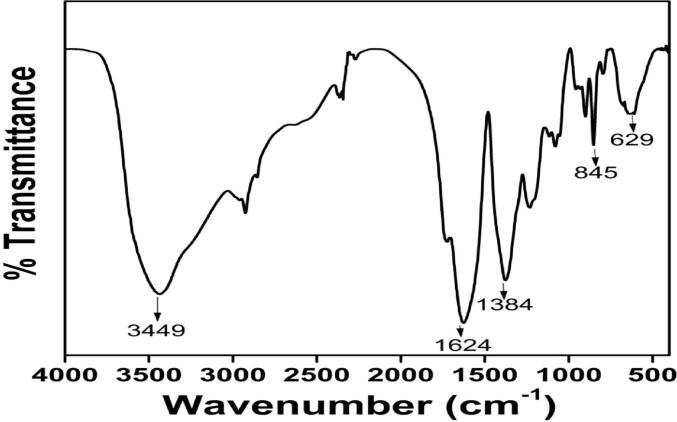
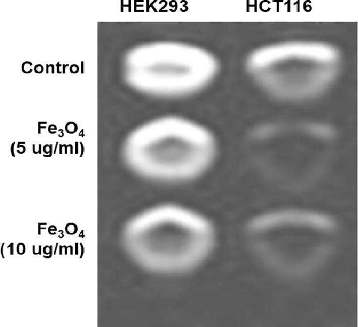
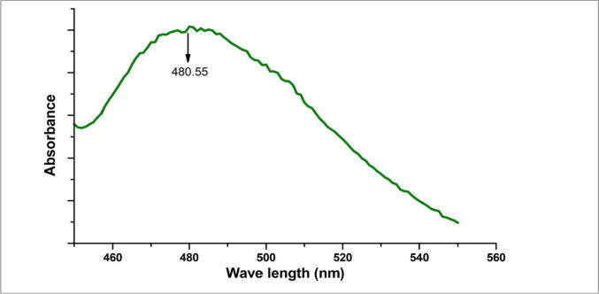
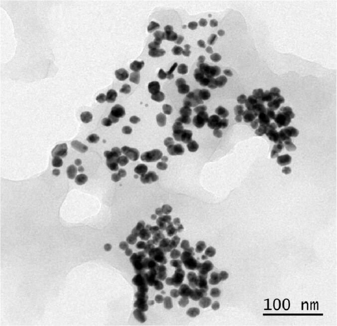
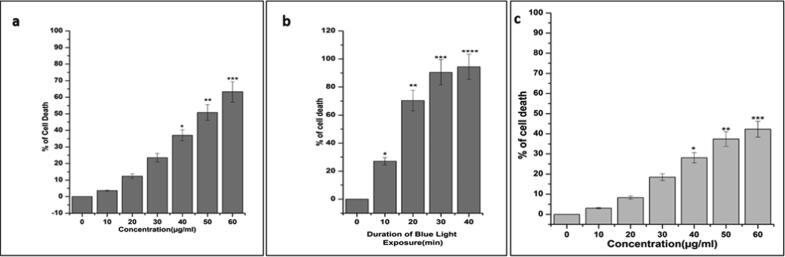
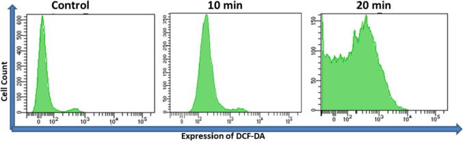
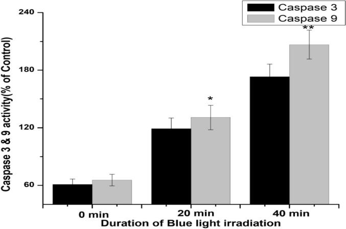
Iron oxides have become increasingly popular for their use as a diagnostic and therapeutic tool in oncology. This study aimed to improve pharmacological valuable of FeO, which may be use to diagnosis colorectal cancers (CRC). Here, we have developed chitosan (CS) coated FeO through a cost-effective procedure. First, we determined the characterization of OA-C-FeO by FTIR, UV-Vis spectra, and TEM. Then, we evaluated the photodynamic therapeutic (PDT) activity of OA-C-FeO in human colorectal carcinoma cell lines (HCT 116). Current results revealed that the light-induced enhanced reactive oxygen species (ROS) activity of the nanoparticles (NPs) and caused cell death the activity of caspase 9/3. The magnetic resonance imaging (MRI) experiments in (HCT 116) and human embryonic kidney cells (HEK 293) illustrated that nanohybrid is an effective MRI contrasting agents for the diagnosis of colorectal cancer.
氧化铁作为肿瘤学中的诊断和治疗工具越来越受欢迎。本研究旨在提高FeO的药理学价值,其可用于诊断结直肠癌(CRC)。在此,我们通过一种经济高效的方法制备了壳聚糖(CS)包覆的FeO。首先,我们通过傅里叶变换红外光谱(FTIR)、紫外可见光谱和透射电子显微镜(TEM)确定了OA-C-FeO的特性。然后,我们评估了OA-C-FeO在人结肠癌细胞系(HCT 116)中的光动力治疗(PDT)活性。目前的结果表明,光诱导纳米颗粒(NPs)的活性氧(ROS)活性增强,并导致细胞死亡以及半胱天冬酶9/3的活性。在(HCT 116)和人胚肾细胞(HEK 293)中进行的磁共振成像(MRI)实验表明,该纳米杂化物是用于诊断结直肠癌的有效MRI造影剂。