文献检索文档翻译深度研究
Suppr Zotero 插件Zotero 插件
邀请有礼套餐&价格历史记录

新学期,新优惠

限时优惠:9月1日-9月22日

30天高级会员仅需29元

1天体验卡首发特惠仅需5.99元

了解详情
不再提醒
插件&应用
Suppr Zotero 插件Zotero 插件浏览器插件Mac 客户端Windows 客户端微信小程序
高级版
套餐订阅购买积分包
AI 工具
文献检索文档翻译深度研究
关于我们
关于 Suppr公司介绍联系我们用户协议隐私条款
关注我们

Suppr 超能文献

核心技术专利:CN118964589B侵权必究
粤ICP备2023148730 号-1Suppr @ 2025

Gouty Arthropathy: Review of Clinical Manifestations and Treatment, with Emphasis on Imaging.

作者信息

Weaver Jennifer S, Vina Ernest R, Munk Peter L, Klauser Andrea S, Elifritz Jamie M, Taljanovic Mihra S

机构信息

Department of Radiology, University of New Mexico, Albuquerque, NM 87131, USA.

Department of Medicine, University of Arizona Arthritis Center, Tucson, AZ 85724, USA.

出版信息

J Clin Med. 2021 Dec 29;11(1):166. doi: 10.3390/jcm11010166.


DOI:10.3390/jcm11010166
PMID:35011907
原文链接:https://pmc.ncbi.nlm.nih.gov/articles/PMC8745871/
Abstract

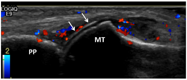
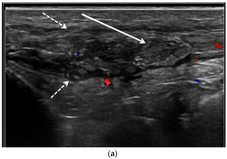
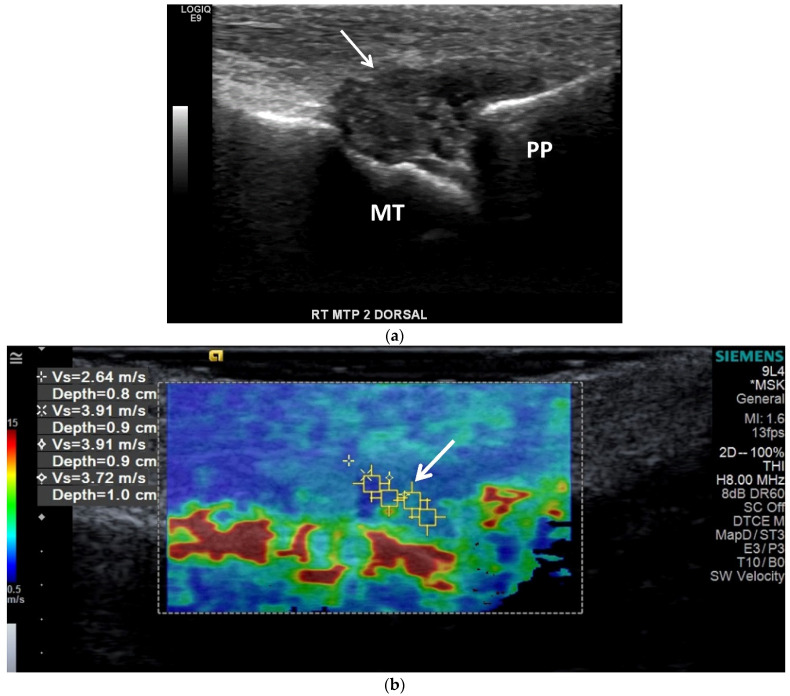
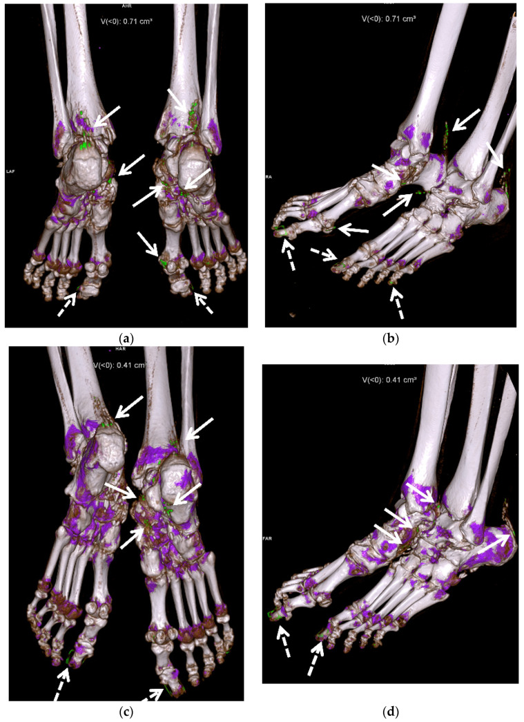
Gout, a crystalline arthropathy caused by the deposition of monosodium urate crystals in the articular and periarticular soft tissues, is a frequent cause of painful arthropathy. Imaging has an important role in the initial evaluation as well as the treatment and follow up of gouty arthropathy. The imaging findings of gouty arthropathy on radiography, ultrasonography, computed tomography, dual energy computed tomography, and magnetic resonance imaging are described to include findings of the early, acute and chronic phases of gout. These findings include early monosodium urate deposits, osseous erosions, and tophi, which may involve periarticular tissues, tendons, and bursae. Treatment of gout includes non-steroidal anti-inflammatories, colchicine, glucocorticoids, interleukin-1 inhibitors, xanthine oxidase inhibitors, uricosuric drugs, and recombinant uricase. Imaging is critical in monitoring response to therapy; clinical management can be modulated based on imaging findings. This review article describes the current standard of care in imaging and treatment of gouty arthropathy.

摘要
https://cdn.ncbi.nlm.nih.gov/pmc/blobs/009b/8745871/7210da2fcbc6/jcm-11-00166-g014.jpg
https://cdn.ncbi.nlm.nih.gov/pmc/blobs/009b/8745871/3e6ca1225130/jcm-11-00166-g001.jpg
https://cdn.ncbi.nlm.nih.gov/pmc/blobs/009b/8745871/a56fcacaa4da/jcm-11-00166-g002.jpg
https://cdn.ncbi.nlm.nih.gov/pmc/blobs/009b/8745871/f202345cc2ad/jcm-11-00166-g003.jpg
https://cdn.ncbi.nlm.nih.gov/pmc/blobs/009b/8745871/3b0f271eee4d/jcm-11-00166-g004a.jpg
https://cdn.ncbi.nlm.nih.gov/pmc/blobs/009b/8745871/f25757b5bfb9/jcm-11-00166-g005.jpg
https://cdn.ncbi.nlm.nih.gov/pmc/blobs/009b/8745871/ad72a1134186/jcm-11-00166-g006.jpg
https://cdn.ncbi.nlm.nih.gov/pmc/blobs/009b/8745871/664241b38ce8/jcm-11-00166-g007.jpg
https://cdn.ncbi.nlm.nih.gov/pmc/blobs/009b/8745871/f1a7dfd641d5/jcm-11-00166-g008.jpg
https://cdn.ncbi.nlm.nih.gov/pmc/blobs/009b/8745871/d53643a9fcaf/jcm-11-00166-g009.jpg
https://cdn.ncbi.nlm.nih.gov/pmc/blobs/009b/8745871/83afb2d16f86/jcm-11-00166-g010a.jpg
https://cdn.ncbi.nlm.nih.gov/pmc/blobs/009b/8745871/a11a41f2641d/jcm-11-00166-g011.jpg
https://cdn.ncbi.nlm.nih.gov/pmc/blobs/009b/8745871/e455da8d7484/jcm-11-00166-g012.jpg
https://cdn.ncbi.nlm.nih.gov/pmc/blobs/009b/8745871/715d85fd4883/jcm-11-00166-g013.jpg
https://cdn.ncbi.nlm.nih.gov/pmc/blobs/009b/8745871/7210da2fcbc6/jcm-11-00166-g014.jpg

相似文献

[1]
Gouty Arthropathy: Review of Clinical Manifestations and Treatment, with Emphasis on Imaging.

J Clin Med. 2021-12-29

[2]
Gouty arthropathy: Review of clinico-pathologic and imaging features.

J Med Imaging Radiat Oncol. 2016-2

[3]
Urate Crystals; Beyond Joints.

Front Med (Lausanne). 2021-6-4

[4]
Tophaceous Gout in an Anorectic Patient Visualized by Dual Energy Computed Tomography (DECT).

Am J Case Rep. 2016-7-15

[5]
Dual-Energy Computed Tomography of the Knee, Ankle, and Foot: Noninvasive Diagnosis of Gout and Quantification of Monosodium Urate in Tendons and Ligaments.

Semin Musculoskelet Radiol. 2016-2

[6]
Imaging in gout--what can we learn from MRI, CT, DECT and US?

Arthritis Res Ther. 2011-11-4

[7]
Imaging of gout: findings and pitfalls. A pictorial review.

Acta Reumatol Port. 2020

[8]
Fine-needle aspiration cytology of periarticular nodule - Atypical presentation of gouty tophi: A report of two cases.

Cytojournal. 2022-8-5

[9]
Computed Tomography and MR Imaging in Crystalline-Induced Arthropathies.

Radiol Clin North Am. 2017-9

[10]
Dual-Energy Computed Tomography (DECT) Resolves the Diagnostic Dilemma in an Atypically Presenting Case of Gout.

Cureus. 2023-4-28

引用本文的文献

[1]
Clinical observations on arthroscopic removal of urate deposits in the knee: a seven-case series.

Eur J Orthop Surg Traumatol. 2025-6-23

[2]
Imaging of gout: an atlas.

Rheumatol Adv Pract. 2025-6-4

[3]
Case Report: Sliding gouty tophi: a case of dynamic ultrasound diagnosis of carpal tunnel syndrome.

Front Surg. 2025-5-13

[4]
Total Knee Arthroplasty for Severe Crystalline-Induced Arthropathy: A Case Report From a Third-Level Hospital.

Cureus. 2025-4-17

[5]
Auricular Gouty Tophi: A Rare Presentation in an Uncommon Site.

Cureus. 2025-3-23

[6]
Deciphering the role of cell signaling pathways in gout pathogenesis and the therapeutic potential of phytoconstituents in their modulation.

Inflammopharmacology. 2025-4-18

[7]
Pictorial Review of Soft Tissue Lesions with Calcification.

Diagnostics (Basel). 2025-3-22

[8]
Risk of Gout Among Patients With Tuberculosis: A Nationwide Cohort Study in South Korea.

Int J Rheum Dis. 2025-3

[9]
Tophaceous Gout in the Axial Skeleton: An Unusual Case with Key Imaging Characteristics.

Kans J Med. 2025-2-17

[10]
Association between advanced lung cancer inflammation index and acute gouty arthritis in Dalian, China: a cross-sectional study.

Front Nutr. 2025-2-14

本文引用的文献

[1]
The role of dual energy computed tomography in the differentiation of acute gout flares and acute calcium pyrophosphate crystal arthritis.

Clin Rheumatol. 2022-1

[2]
Pulsed Electromagnetic Fields in Bone Healing: Molecular Pathways and Clinical Applications.

Int J Mol Sci. 2021-7-9

[3]
Two-year reduction of dual-energy CT urate depositions during a treat-to-target strategy in gout in the NOR-Gout longitudinal study.

Rheumatology (Oxford). 2022-4-18

[4]
Sonographic estimation of monosodium urate burden predicts the fulfillment of the 2016 remission criteria for gout: a 12-month study.

Arthritis Res Ther. 2021-7-9

[5]
A Randomized, Phase II Study Evaluating the Efficacy and Safety of Anakinra in the Treatment of Gout Flares.

Arthritis Rheumatol. 2021-8

[6]
Consensus-based semi-quantitative ultrasound scoring system for gout lesions: Results of an OMERACT Delphi process and web-reliability exercise.

Semin Arthritis Rheum. 2021-6

[7]
Crystal deposition measured with dual-energy computed tomography: association with mortality and cardiovascular risks in gout.

Rheumatology (Oxford). 2021-10-2

[8]
Tophaceous Gout.

Ann Acad Med Singap. 2020-11

[9]
Ultrasonography and dual-energy computed tomography: impact for the detection of gouty deposits.

Ultrasonography. 2021-4

[10]
UltraSound evaluation in follow-up of urate-lowering therapy in gout phase 2 (USEFUL-2): Duration of flare prophylaxis.

Joint Bone Spine. 2020-12

文献AI研究员

20分钟写一篇综述,助力文献阅读效率提升50倍

立即体验

用中文搜PubMed

大模型驱动的PubMed中文搜索引擎

马上搜索

推荐工具

医学文档翻译智能文献检索