• 文献检索
  • 文档翻译
  • 深度研究
  • 学术资讯
  • Suppr Zotero 插件Zotero 插件
  • 邀请有礼
  • 套餐&价格
  • 历史记录
应用&插件
Suppr Zotero 插件Zotero 插件浏览器插件Mac 客户端Windows 客户端微信小程序
定价
高级版会员购买积分包购买API积分包
服务
文献检索文档翻译深度研究API 文档MCP 服务
关于我们
关于 Suppr公司介绍联系我们用户协议隐私条款
关注我们

Suppr 超能文献

核心技术专利:CN118964589B侵权必究
粤ICP备2023148730 号-1Suppr @ 2026

文献检索

告别复杂PubMed语法,用中文像聊天一样搜索,搜遍4000万医学文献。AI智能推荐,让科研检索更轻松。

立即免费搜索

文件翻译

保留排版,准确专业,支持PDF/Word/PPT等文件格式,支持 12+语言互译。

免费翻译文档

深度研究

AI帮你快速写综述,25分钟生成高质量综述,智能提取关键信息,辅助科研写作。

立即免费体验

用于数字病理学的深度分割模型的无代码开发与部署

Code-Free Development and Deployment of Deep Segmentation Models for Digital Pathology.

作者信息

Pettersen Henrik Sahlin, Belevich Ilya, Røyset Elin Synnøve, Smistad Erik, Simpson Melanie Rae, Jokitalo Eija, Reinertsen Ingerid, Bakke Ingunn, Pedersen André

机构信息

Department of Pathology, St. Olavs Hospital, Trondheim University Hospital, Trondheim, Norway.

Department of Clinical and Molecular Medicine, Faculty of Medicine and Health Sciences, NTNU - Norwegian University of Science and Technology, Trondheim, Norway.

出版信息

Front Med (Lausanne). 2022 Jan 27;8:816281. doi: 10.3389/fmed.2021.816281. eCollection 2021.

DOI:10.3389/fmed.2021.816281
PMID:35155486
原文链接:https://pmc.ncbi.nlm.nih.gov/articles/PMC8829033/
Abstract

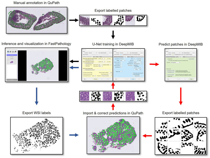
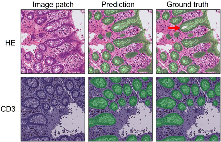
Application of deep learning on histopathological whole slide images (WSIs) holds promise of improving diagnostic efficiency and reproducibility but is largely dependent on the ability to write computer code or purchase commercial solutions. We present a code-free pipeline utilizing free-to-use, open-source software (QuPath, DeepMIB, and FastPathology) for creating and deploying deep learning-based segmentation models for computational pathology. We demonstrate the pipeline on a use case of separating epithelium from stroma in colonic mucosa. A dataset of 251 annotated WSIs, comprising 140 hematoxylin-eosin (HE)-stained and 111 CD3 immunostained colon biopsy WSIs, were developed through active learning using the pipeline. On a hold-out test set of 36 HE and 21 CD3-stained WSIs a mean intersection over union score of 95.5 and 95.3% was achieved on epithelium segmentation. We demonstrate pathologist-level segmentation accuracy and clinical acceptable runtime performance and show that pathologists without programming experience can create near state-of-the-art segmentation solutions for histopathological WSIs using only free-to-use software. The study further demonstrates the strength of open-source solutions in its ability to create generalizable, open pipelines, of which trained models and predictions can seamlessly be exported in open formats and thereby used in external solutions. All scripts, trained models, a video tutorial, and the full dataset of 251 WSIs with ~31 k epithelium annotations are made openly available at https://github.com/andreped/NoCodeSeg to accelerate research in the field.

摘要

将深度学习应用于组织病理学全切片图像(WSIs)有望提高诊断效率和可重复性,但很大程度上依赖于编写计算机代码或购买商业解决方案的能力。我们提出了一种无需代码的流程,利用免费的开源软件(QuPath、DeepMIB和FastPathology)来创建和部署基于深度学习的计算病理学分割模型。我们在结肠黏膜上皮与基质分离的用例中展示了该流程。通过使用该流程进行主动学习,开发了一个包含251个带注释的WSIs的数据集,其中包括140个苏木精-伊红(HE)染色和111个CD3免疫染色的结肠活检WSIs。在36个HE染色和21个CD3染色的WSIs的保留测试集上,上皮分割的平均交并比分数分别达到了95.5%和95.3%。我们展示了病理学家级别的分割准确性和临床可接受的运行时性能,并表明没有编程经验的病理学家仅使用免费软件就能为组织病理学WSIs创建近乎最先进的分割解决方案。该研究进一步证明了开源解决方案在创建可推广的开放流程方面的优势,其中训练好的模型和预测结果可以以开放格式无缝导出,从而用于外部解决方案。所有脚本、训练好的模型、视频教程以及包含约31k上皮注释的251个WSIs的完整数据集可在https://github.com/andreped/NoCodeSeg上公开获取,以加速该领域的研究。

https://cdn.ncbi.nlm.nih.gov/pmc/blobs/1150/8829033/95b98d9de4bc/fmed-08-816281-g0004.jpg
https://cdn.ncbi.nlm.nih.gov/pmc/blobs/1150/8829033/dfd5ecaf6d11/fmed-08-816281-g0001.jpg
https://cdn.ncbi.nlm.nih.gov/pmc/blobs/1150/8829033/ea918a7719be/fmed-08-816281-g0002.jpg
https://cdn.ncbi.nlm.nih.gov/pmc/blobs/1150/8829033/f90bea2a9d5a/fmed-08-816281-g0003.jpg
https://cdn.ncbi.nlm.nih.gov/pmc/blobs/1150/8829033/95b98d9de4bc/fmed-08-816281-g0004.jpg

相似文献

1
Code-Free Development and Deployment of Deep Segmentation Models for Digital Pathology.用于数字病理学的深度分割模型的无代码开发与部署
Front Med (Lausanne). 2022 Jan 27;8:816281. doi: 10.3389/fmed.2021.816281. eCollection 2021.
2
Deep learning for colon cancer histopathological images analysis.用于结肠癌组织病理学图像分析的深度学习
Comput Biol Med. 2021 Sep;136:104730. doi: 10.1016/j.compbiomed.2021.104730. Epub 2021 Aug 4.
3
Equipping computational pathology systems with artifact processing pipelines: a showcase for computation and performance trade-offs.为计算病理学系统配备伪影处理管道:计算和性能权衡的展示。
BMC Med Inform Decis Mak. 2024 Oct 7;24(1):288. doi: 10.1186/s12911-024-02676-z.
4
Automated diagnosis of 7 canine skin tumors using machine learning on H&E-stained whole slide images.基于 H&E 染色全切片图像的机器学习对 7 例犬皮肤肿瘤的自动诊断
Vet Pathol. 2023 Nov;60(6):865-875. doi: 10.1177/03009858231189205. Epub 2023 Jul 29.
5
Deep learning-based image analysis methods for brightfield-acquired multiplex immunohistochemistry images.基于深度学习的明场获取多重免疫组化图像的图像分析方法。
Diagn Pathol. 2020 Jul 28;15(1):100. doi: 10.1186/s13000-020-01003-0.
6
A U-Net based framework to quantify glomerulosclerosis in digitized PAS and H&E stained human tissues.基于 U-Net 的框架来量化数字化 PAS 和 H&E 染色的人类组织中的肾小球硬化。
Comput Med Imaging Graph. 2021 Apr;89:101865. doi: 10.1016/j.compmedimag.2021.101865. Epub 2021 Jan 28.
7
Deep learning-based semantic segmentation of non-melanocytic skin tumors in whole-slide histopathological images.基于深度学习的全切片组织病理图像中非黑色素性皮肤肿瘤的语义分割。
Exp Dermatol. 2023 Jun;32(6):831-839. doi: 10.1111/exd.14782. Epub 2023 Apr 5.
8
Lung Cancer Diagnosis on Virtual Histologically Stained Tissue Using Weakly Supervised Learning.基于弱监督学习的虚拟组织染色切片肺癌诊断
Mod Pathol. 2024 Jun;37(6):100487. doi: 10.1016/j.modpat.2024.100487. Epub 2024 Apr 7.
9
An Accelerated Pipeline for Multi-label Renal Pathology Image Segmentation at the Whole Slide Image Level.一种用于全切片图像级多标签肾脏病理图像分割的加速流程
Proc SPIE Int Soc Opt Eng. 2023 Feb;12471. doi: 10.1117/12.2653651. Epub 2023 Apr 6.
10
HoLy-Net: Segmentation of histological images of diffuse large B-cell lymphoma.HoLy-Net:弥漫性大 B 细胞淋巴瘤组织学图像分割。
Comput Biol Med. 2024 Mar;170:107978. doi: 10.1016/j.compbiomed.2024.107978. Epub 2024 Jan 11.

引用本文的文献

1
Leveraging pre-trained machine learning models for islet quantification in type 1 diabetes.利用预训练机器学习模型进行1型糖尿病中的胰岛定量分析。
J Pathol Inform. 2024 Nov 8;16:100406. doi: 10.1016/j.jpi.2024.100406. eCollection 2025 Jan.
2
Development of an Artificial-Intelligence-Based Tool for Automated Assessment of Cellularity in Bone Marrow Biopsies in Ph-Negative Myeloproliferative Neoplasms.基于人工智能的阴性骨髓增殖性肿瘤骨髓活检细胞密度自动评估工具的开发
Cancers (Basel). 2024 Apr 26;16(9):1687. doi: 10.3390/cancers16091687.
3
tRigon: an R package and Shiny App for integrative (path-)omics data analysis.

本文引用的文献

1
Quick Annotator: an open-source digital pathology based rapid image annotation tool.快速标注器:一个基于开源数字病理学的快速图像标注工具。
J Pathol Clin Res. 2021 Nov;7(6):542-547. doi: 10.1002/cjp2.229. Epub 2021 Jul 19.
2
Deep learning in histopathology: the path to the clinic.深度学习在组织病理学中的应用:通往临床的道路。
Nat Med. 2021 May;27(5):775-784. doi: 10.1038/s41591-021-01343-4. Epub 2021 May 14.
3
Democratising deep learning for microscopy with ZeroCostDL4Mic.使用 ZeroCostDL4Mic 实现显微镜深度学习民主化。
tRigon:一个用于整合(路径)组学数据分析的 R 包和 Shiny 应用程序。
BMC Bioinformatics. 2024 Mar 5;25(1):98. doi: 10.1186/s12859-024-05721-w.
4
Sex Differences in Glomerular Lesions, in Atherosclerosis Progression, and in the Response to Angiotensin-Converting Enzyme Inhibitors in the ApoE Mice Model.载脂蛋白 E 基因敲除小鼠模型中肾小球病变、动脉粥样硬化进展和血管紧张素转换酶抑制剂反应的性别差异。
Int J Mol Sci. 2023 Aug 30;24(17):13442. doi: 10.3390/ijms241713442.
5
Deep learning-based image analysis reveals significant differences in the number and distribution of mucosal CD3 and γδ T cells between Crohn's disease and ulcerative colitis.基于深度学习的图像分析揭示了克罗恩病和溃疡性结肠炎之间黏膜 CD3 和 γδ T 细胞数量和分布的显著差异。
J Pathol Clin Res. 2023 Jan;9(1):18-31. doi: 10.1002/cjp2.301. Epub 2022 Nov 23.
Nat Commun. 2021 Apr 15;12(1):2276. doi: 10.1038/s41467-021-22518-0.
4
Quantitative Image Analysis for Tissue Biomarker Use: A White Paper From the Digital Pathology Association.用于组织生物标志物的定量图像分析:数字病理协会白皮书
Appl Immunohistochem Mol Morphol. 2021 Aug 1;29(7):479-493. doi: 10.1097/PAI.0000000000000930.
5
DeepMIB: User-friendly and open-source software for training of deep learning network for biological image segmentation.DeepMIB:用于生物图像分割的深度学习网络训练的用户友好型和开源软件。
PLoS Comput Biol. 2021 Mar 2;17(3):e1008374. doi: 10.1371/journal.pcbi.1008374. eCollection 2021 Mar.
6
nnU-Net: a self-configuring method for deep learning-based biomedical image segmentation.nnU-Net:一种基于深度学习的生物医学图像分割的自配置方法。
Nat Methods. 2021 Feb;18(2):203-211. doi: 10.1038/s41592-020-01008-z. Epub 2020 Dec 7.
7
Digital Pathology: Advantages, Limitations and Emerging Perspectives.数字病理学:优势、局限性与新兴观点
J Clin Med. 2020 Nov 18;9(11):3697. doi: 10.3390/jcm9113697.
8
Deep neural network models for computational histopathology: A survey.用于计算组织病理学的深度神经网络模型:一项综述。
Med Image Anal. 2021 Jan;67:101813. doi: 10.1016/j.media.2020.101813. Epub 2020 Sep 25.
9
Automated deep-learning system for Gleason grading of prostate cancer using biopsies: a diagnostic study.利用活检进行前列腺癌 Gleason 分级的自动化深度学习系统:一项诊断研究。
Lancet Oncol. 2020 Feb;21(2):233-241. doi: 10.1016/S1470-2045(19)30739-9. Epub 2020 Jan 8.
10
An integrated iterative annotation technique for easing neural network training in medical image analysis.一种用于简化医学图像分析中神经网络训练的集成迭代标注技术。
Nat Mach Intell. 2019 Feb;1(2):112-119. doi: 10.1038/s42256-019-0018-3. Epub 2019 Feb 11.