• 文献检索
  • 文档翻译
  • 深度研究
  • 学术资讯
  • Suppr Zotero 插件Zotero 插件
  • 邀请有礼
  • 套餐&价格
  • 历史记录
应用&插件
Suppr Zotero 插件Zotero 插件浏览器插件Mac 客户端Windows 客户端微信小程序
定价
高级版会员购买积分包购买API积分包
服务
文献检索文档翻译深度研究API 文档MCP 服务
关于我们
关于 Suppr公司介绍联系我们用户协议隐私条款
关注我们

Suppr 超能文献

核心技术专利:CN118964589B侵权必究
粤ICP备2023148730 号-1Suppr @ 2026

文献检索

告别复杂PubMed语法,用中文像聊天一样搜索,搜遍4000万医学文献。AI智能推荐,让科研检索更轻松。

立即免费搜索

文件翻译

保留排版,准确专业,支持PDF/Word/PPT等文件格式,支持 12+语言互译。

免费翻译文档

深度研究

AI帮你快速写综述,25分钟生成高质量综述,智能提取关键信息,辅助科研写作。

立即免费体验

快速标注器:一个基于开源数字病理学的快速图像标注工具。

Quick Annotator: an open-source digital pathology based rapid image annotation tool.

机构信息

Department of Biomedical Engineering, Case Western Reserve University, Cleveland, OH, USA.

Toth Technology LLC, Dover, NJ, USA.

出版信息

J Pathol Clin Res. 2021 Nov;7(6):542-547. doi: 10.1002/cjp2.229. Epub 2021 Jul 19.

DOI:10.1002/cjp2.229
PMID:34288586
原文链接:https://pmc.ncbi.nlm.nih.gov/articles/PMC8503896/
Abstract

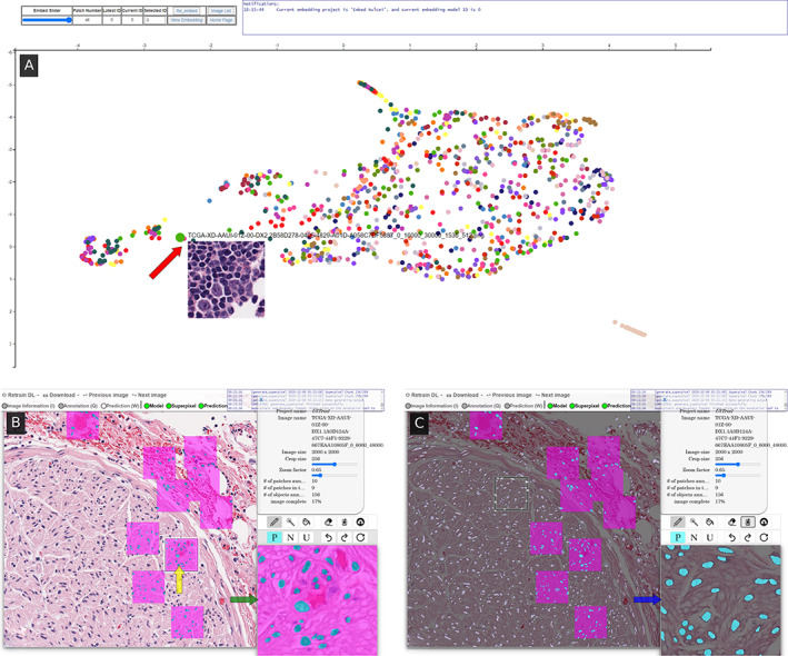
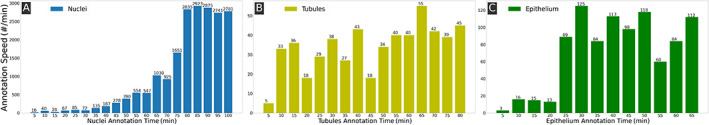
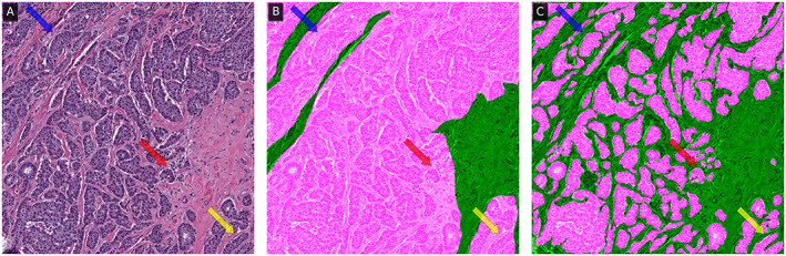
Image-based biomarker discovery typically requires accurate segmentation of histologic structures (e.g. cell nuclei, tubules, and epithelial regions) in digital pathology whole slide images (WSIs). Unfortunately, annotating each structure of interest is laborious and often intractable even in moderately sized cohorts. Here, we present an open-source tool, Quick Annotator (QA), designed to improve annotation efficiency of histologic structures by orders of magnitude. While the user annotates regions of interest (ROIs) via an intuitive web interface, a deep learning (DL) model is concurrently optimized using these annotations and applied to the ROI. The user iteratively reviews DL results to either (1) accept accurately annotated regions or (2) correct erroneously segmented structures to improve subsequent model suggestions, before transitioning to other ROIs. We demonstrate the effectiveness of QA over comparable manual efforts via three use cases. These include annotating (1) 337,386 nuclei in 5 pancreatic WSIs, (2) 5,692 tubules in 10 colorectal WSIs, and (3) 14,187 regions of epithelium in 10 breast WSIs. Efficiency gains in terms of annotations per second of 102×, 9×, and 39× were, respectively, witnessed while retaining f-scores >0.95, suggesting that QA may be a valuable tool for efficiently fully annotating WSIs employed in downstream biomarker studies.

摘要

基于图像的生物标志物发现通常需要在数字病理学全切片图像 (WSI) 中准确分割组织学结构(例如细胞核、小管和上皮区域)。不幸的是,即使在中等大小的队列中,注释每个感兴趣的结构也是费力且通常难以解决的。在这里,我们提出了一个开源工具,即快速注释器 (Quick Annotator,QA),旨在通过数量级提高组织学结构注释的效率。当用户通过直观的 Web 界面注释感兴趣的区域 (ROI) 时,一个深度学习 (DL) 模型会同时使用这些注释进行优化,并应用于 ROI。用户会反复查看 DL 结果,要么 (1) 接受准确注释的区域,要么 (2) 纠正错误分割的结构,以提高后续模型建议的准确性,然后再转到其他 ROI。我们通过三个用例证明了 QA 相对于可比手动工作的有效性。这些用例包括注释 (1) 5 个胰腺 WSI 中的 337386 个细胞核、(2) 10 个结直肠 WSI 中的 5692 个小管和 (3) 10 个乳腺 WSI 中的 14187 个上皮区域。在保留 f-score>0.95 的情况下,分别见证了每秒注释数提高了 102×、9× 和 39×的效率增益,这表明 QA 可能是一种非常有价值的工具,可用于高效地完全注释用于下游生物标志物研究的 WSI。

https://cdn.ncbi.nlm.nih.gov/pmc/blobs/5f20/8503896/7c62fae8aab4/CJP2-7-542-g002.jpg
https://cdn.ncbi.nlm.nih.gov/pmc/blobs/5f20/8503896/726e8afa9d50/CJP2-7-542-g003.jpg
https://cdn.ncbi.nlm.nih.gov/pmc/blobs/5f20/8503896/b4e996fd12ad/CJP2-7-542-g001.jpg
https://cdn.ncbi.nlm.nih.gov/pmc/blobs/5f20/8503896/7c62fae8aab4/CJP2-7-542-g002.jpg
https://cdn.ncbi.nlm.nih.gov/pmc/blobs/5f20/8503896/726e8afa9d50/CJP2-7-542-g003.jpg
https://cdn.ncbi.nlm.nih.gov/pmc/blobs/5f20/8503896/b4e996fd12ad/CJP2-7-542-g001.jpg
https://cdn.ncbi.nlm.nih.gov/pmc/blobs/5f20/8503896/7c62fae8aab4/CJP2-7-542-g002.jpg

相似文献

1
Quick Annotator: an open-source digital pathology based rapid image annotation tool.快速标注器:一个基于开源数字病理学的快速图像标注工具。
J Pathol Clin Res. 2021 Nov;7(6):542-547. doi: 10.1002/cjp2.229. Epub 2021 Jul 19.
2
From whole-slide image to biomarker prediction: end-to-end weakly supervised deep learning in computational pathology.从全切片图像到生物标志物预测:计算病理学中的端到端弱监督深度学习
Nat Protoc. 2025 Jan;20(1):293-316. doi: 10.1038/s41596-024-01047-2. Epub 2024 Sep 16.
3
Assessment of a computerized quantitative quality control tool for whole slide images of kidney biopsies.用于肾活检全切片图像的计算机化定量质量控制工具的评估。
J Pathol. 2021 Mar;253(3):268-278. doi: 10.1002/path.5590. Epub 2021 Jan 5.
4
Development and clinical validation of deep learning-based immunohistochemistry prediction models for subtyping and staging of gastrointestinal cancers.基于深度学习的胃肠道癌亚型和分期免疫组织化学预测模型的开发与临床验证
BMC Gastroenterol. 2025 Jul 1;25(1):494. doi: 10.1186/s12876-025-04045-0.
5
Variation within and between digital pathology and light microscopy for the diagnosis of histopathology slides: blinded crossover comparison study.数字病理学与光学显微镜检查在组织病理学切片诊断中的内部及相互间差异:双盲交叉对比研究
Health Technol Assess. 2025 Jul;29(30):1-75. doi: 10.3310/SPLK4325.
6
Exploratory multi-cohort, multi-reader study on the clinical utility of a deep learning model for transforming cryosectioned to formalin-fixed, paraffin-embedded (FFPE) images in breast lesion diagnosis.关于深度学习模型在乳腺病变诊断中用于将冷冻切片图像转换为福尔马林固定石蜡包埋(FFPE)图像的临床效用的探索性多队列、多读者研究。
Breast Cancer Res. 2025 Jun 17;27(1):110. doi: 10.1186/s13058-025-02064-z.
7
An open-source platform for structured annotation and computational workflows in digital pathology research.一个用于数字病理学研究中结构化注释和计算工作流程的开源平台。
Sci Rep. 2025 Aug 7;15(1):28910. doi: 10.1038/s41598-025-13546-7.
8
Self-supervised deep metric learning for prototypical zero-shot lesion retrieval in placenta whole-slide images.用于胎盘全切片图像中原型零样本病变检索的自监督深度度量学习
Comput Biol Med. 2025 Sep;196(Pt A):110634. doi: 10.1016/j.compbiomed.2025.110634. Epub 2025 Jul 3.
9
Labor-Efficient Pathological Auxiliary Diagnostic Model for Primary and Metastatic Tumor Tissue Detection in Pancreatic Ductal Adenocarcinoma.用于胰腺导管腺癌原发和转移瘤组织检测的劳动高效型病理辅助诊断模型
Mod Pathol. 2025 Jul;38(7):100764. doi: 10.1016/j.modpat.2025.100764. Epub 2025 Apr 6.
10
Tumour purity assessment with deep learning in colorectal cancer and impact on molecular analysis.利用深度学习评估结直肠癌肿瘤纯度及其对分子分析的影响
J Pathol. 2025 Feb;265(2):184-197. doi: 10.1002/path.6376. Epub 2024 Dec 22.

引用本文的文献

1
Development and clinical validation of deep learning-based immunohistochemistry prediction models for subtyping and staging of gastrointestinal cancers.基于深度学习的胃肠道癌亚型和分期免疫组织化学预测模型的开发与临床验证
BMC Gastroenterol. 2025 Jul 1;25(1):494. doi: 10.1186/s12876-025-04045-0.
2
Machine Vision-Detected Peritumoral Lymphocytic Aggregates Are Associated With Disease-Free Survival in Patients With Papillary Thyroid Carcinoma.机器视觉检测到的甲状腺乳头状癌患者瘤周淋巴细胞聚集与无病生存期相关。
Lab Invest. 2024 Dec;104(12):102168. doi: 10.1016/j.labinv.2024.102168. Epub 2024 Nov 4.
3
Improving the Annotation Process in Computational Pathology: A Pilot Study with Manual and Semi-automated Approaches on Consumer and Medical Grade Devices.

本文引用的文献

1
Feature-driven local cell graph (FLocK): New computational pathology-based descriptors for prognosis of lung cancer and HPV status of oropharyngeal cancers.特征驱动的局部细胞图谱(FLocK):基于计算病理学的肺癌预后及口咽癌HPV状态的新描述符
Med Image Anal. 2021 Feb;68:101903. doi: 10.1016/j.media.2020.101903. Epub 2020 Nov 16.
2
Reducing annotation effort in digital pathology: A Co-Representation learning framework for classification tasks.减少数字病理学中的注释工作:用于分类任务的协同表示学习框架。
Med Image Anal. 2021 Jan;67:101859. doi: 10.1016/j.media.2020.101859. Epub 2020 Oct 9.
3
TissueWand, a Rapid Histopathology Annotation Tool.
改进计算病理学中的标注过程:一项关于消费级和医疗级设备的手动及半自动方法的试点研究。
J Imaging Inform Med. 2025 Apr;38(2):1112-1119. doi: 10.1007/s10278-024-01248-x. Epub 2024 Sep 4.
4
Computational methods and biomarker discovery strategies for spatial proteomics: a review in immuno-oncology.计算方法和生物标志物发现策略在空间蛋白质组学中的应用:免疫肿瘤学综述。
Brief Bioinform. 2024 Jul 25;25(5). doi: 10.1093/bib/bbae421.
5
Enhancing AI Research for Breast Cancer: A Comprehensive Review of Tumor-Infiltrating Lymphocyte Datasets.加强乳腺癌的人工智能研究:肿瘤浸润淋巴细胞数据集的综合综述
J Imaging Inform Med. 2024 Dec;37(6):2996-3008. doi: 10.1007/s10278-024-01043-8. Epub 2024 May 28.
6
Closing the loop - the role of pathologists in digital and computational pathology research.闭环——病理学家在数字和计算病理学研究中的作用。
J Pathol Clin Res. 2024 Mar;10(2):e12366. doi: 10.1002/2056-4538.12366.
7
Computational pathology: A survey review and the way forward.计算病理学:综述与未来发展方向
J Pathol Inform. 2024 Jan 14;15:100357. doi: 10.1016/j.jpi.2023.100357. eCollection 2024 Dec.
8
SlideTiler: A dataset creator software for boosting deep learning on histological whole slide images.SlideTiler:一种用于在组织学全切片图像上促进深度学习的数据集创建软件。
J Pathol Inform. 2023 Dec 8;15:100356. doi: 10.1016/j.jpi.2023.100356. eCollection 2024 Dec.
9
Accelerating pharmaceutical R&D with a user-friendly AI system for histopathology image analysis.利用用户友好型人工智能系统进行组织病理学图像分析以加速药物研发。
J Pathol Inform. 2023 Sep 25;14:100337. doi: 10.1016/j.jpi.2023.100337. eCollection 2023.
10
Effective and efficient active learning for deep learning-based tissue image analysis.基于深度学习的组织图像分析的有效和高效主动学习。
Bioinformatics. 2023 Apr 3;39(4). doi: 10.1093/bioinformatics/btad138.
组织棒,一种快速组织病理学标注工具。
J Pathol Inform. 2020 Aug 21;11:27. doi: 10.4103/jpi.jpi_5_20. eCollection 2020.
4
Dataset of segmented nuclei in hematoxylin and eosin stained histopathology images of ten cancer types.十类癌症的苏木精和伊红染色组织病理学图像分割细胞核数据集。
Sci Data. 2020 Jun 19;7(1):185. doi: 10.1038/s41597-020-0528-1.
5
Cellular community detection for tissue phenotyping in colorectal cancer histology images.用于结直肠癌组织学图像组织表型分析的细胞群落检测。
Med Image Anal. 2020 Jul;63:101696. doi: 10.1016/j.media.2020.101696. Epub 2020 Apr 13.
6
Batch Mode Active Learning on the Riemannian Manifold for Automated Scoring of Nuclear Pleomorphism in Breast Cancer.基于黎曼流形的批处理主动学习在乳腺癌核异型性自动评分中的应用。
Artif Intell Med. 2020 Mar;103:101805. doi: 10.1016/j.artmed.2020.101805. Epub 2020 Jan 25.
7
Topological Tumor Graphs: A Graph-Based Spatial Model to Infer Stromal Recruitment for Immunosuppression in Melanoma Histology.拓扑肿瘤图:一种基于图的空间模型,用于推断黑色素瘤组织学中基质募集的免疫抑制作用。
Cancer Res. 2020 Mar 1;80(5):1199-1209. doi: 10.1158/0008-5472.CAN-19-2268. Epub 2019 Dec 24.
8
Strategies to Reduce the Expert Supervision Required for Deep Learning-Based Segmentation of Histopathological Images.减少基于深度学习的组织病理学图像分割所需专家监督的策略
Front Med (Lausanne). 2019 Oct 15;6:222. doi: 10.3389/fmed.2019.00222. eCollection 2019.
9
An integrated iterative annotation technique for easing neural network training in medical image analysis.一种用于简化医学图像分析中神经网络训练的集成迭代标注技术。
Nat Mach Intell. 2019 Feb;1(2):112-119. doi: 10.1038/s42256-019-0018-3. Epub 2019 Feb 11.
10
Quantitative nuclear histomorphometry predicts oncotype DX risk categories for early stage ER+ breast cancer.定量核组织形态计量学可预测早期 ER+ 乳腺癌的 Oncotype DX 风险类别。
BMC Cancer. 2018 May 30;18(1):610. doi: 10.1186/s12885-018-4448-9.