• 文献检索
  • 文档翻译
  • 深度研究
  • 学术资讯
  • Suppr Zotero 插件Zotero 插件
  • 邀请有礼
  • 套餐&价格
  • 历史记录
应用&插件
Suppr Zotero 插件Zotero 插件浏览器插件Mac 客户端Windows 客户端微信小程序
定价
高级版会员购买积分包购买API积分包
服务
文献检索文档翻译深度研究API 文档MCP 服务
关于我们
关于 Suppr公司介绍联系我们用户协议隐私条款
关注我们

Suppr 超能文献

核心技术专利:CN118964589B侵权必究
粤ICP备2023148730 号-1Suppr @ 2026

文献检索

告别复杂PubMed语法,用中文像聊天一样搜索,搜遍4000万医学文献。AI智能推荐,让科研检索更轻松。

立即免费搜索

文件翻译

保留排版,准确专业,支持PDF/Word/PPT等文件格式,支持 12+语言互译。

免费翻译文档

深度研究

AI帮你快速写综述,25分钟生成高质量综述,智能提取关键信息,辅助科研写作。

立即免费体验

使用改进的多任务深度学习模型对食管病变进行多分类和分割诊断。

Diagnosis of Esophageal Lesions by Multi-Classification and Segmentation Using an Improved Multi-Task Deep Learning Model.

机构信息

Faculty of Information Technology, Macau University of Science and Technology, Macau 999078, China.

Kiang Wu Hospital, Macau 999078, China.

出版信息

Sensors (Basel). 2022 Feb 15;22(4):1492. doi: 10.3390/s22041492.

DOI:10.3390/s22041492
PMID:35214396
原文链接:https://pmc.ncbi.nlm.nih.gov/articles/PMC8876234/
Abstract

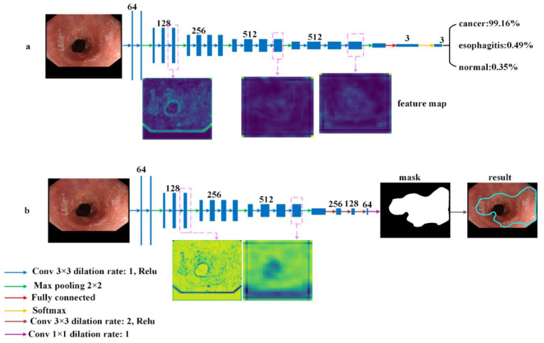
It is challenging for endoscopists to accurately detect esophageal lesions during gastrointestinal endoscopic screening due to visual similarities among different lesions in terms of shape, size, and texture among patients. Additionally, endoscopists are busy fighting esophageal lesions every day, hence the need to develop a computer-aided diagnostic tool to classify and segment the lesions at endoscopic images to reduce their burden. Therefore, we propose a multi-task classification and segmentation (MTCS) model, including the Esophageal Lesions Classification Network (ELCNet) and Esophageal Lesions Segmentation Network (ELSNet). The ELCNet was used to classify types of esophageal lesions, and the ELSNet was used to identify lesion regions. We created a dataset by collecting 805 esophageal images from 255 patients and 198 images from 64 patients to train and evaluate the MTCS model. Compared with other methods, the proposed not only achieved a high accuracy (93.43%) in classification but achieved a dice similarity coefficient (77.84%) in segmentation. In conclusion, the MTCS model can boost the performance of endoscopists in the detection of esophageal lesions as it can accurately multi-classify and segment the lesions and is a potential assistant for endoscopists to reduce the risk of oversight.

摘要

由于患者之间不同病变在形状、大小和纹理等方面存在视觉相似性,内镜医生很难在胃肠内镜筛查中准确检测到食管病变。此外,内镜医生每天都忙于与食管病变作斗争,因此需要开发一种计算机辅助诊断工具,以便对内镜图像中的病变进行分类和分割,以减轻他们的负担。因此,我们提出了一种多任务分类和分割(MTCS)模型,包括食管病变分类网络(ELCNet)和食管病变分割网络(ELSNet)。ELCNet 用于分类食管病变类型,ELSNet 用于识别病变区域。我们通过收集 255 名患者的 805 张食管图像和 64 名患者的 198 张图像来创建数据集,以训练和评估 MTCS 模型。与其他方法相比,该方法不仅在分类方面达到了很高的准确率(93.43%),而且在分割方面达到了较高的骰子相似系数(77.84%)。总之,MTCS 模型可以提高内镜医生检测食管病变的性能,因为它可以准确地对病变进行多分类和分割,是内镜医生减少漏诊风险的潜在辅助手段。

https://cdn.ncbi.nlm.nih.gov/pmc/blobs/fb3b/8876234/3f1d3c8e70c8/sensors-22-01492-g006.jpg
https://cdn.ncbi.nlm.nih.gov/pmc/blobs/fb3b/8876234/8bfb16352768/sensors-22-01492-g001.jpg
https://cdn.ncbi.nlm.nih.gov/pmc/blobs/fb3b/8876234/b75e834051c2/sensors-22-01492-g002.jpg
https://cdn.ncbi.nlm.nih.gov/pmc/blobs/fb3b/8876234/62ba67043119/sensors-22-01492-g003.jpg
https://cdn.ncbi.nlm.nih.gov/pmc/blobs/fb3b/8876234/06eafba654ec/sensors-22-01492-g004.jpg
https://cdn.ncbi.nlm.nih.gov/pmc/blobs/fb3b/8876234/1b0186c0cf93/sensors-22-01492-g005.jpg
https://cdn.ncbi.nlm.nih.gov/pmc/blobs/fb3b/8876234/3f1d3c8e70c8/sensors-22-01492-g006.jpg

相似文献

1
Diagnosis of Esophageal Lesions by Multi-Classification and Segmentation Using an Improved Multi-Task Deep Learning Model.使用改进的多任务深度学习模型对食管病变进行多分类和分割诊断。
Sensors (Basel). 2022 Feb 15;22(4):1492. doi: 10.3390/s22041492.
2
Multi-Task Model for Esophageal Lesion Analysis Using Endoscopic Images: Classification with Image Retrieval and Segmentation with Attention.基于内镜图像的食管病变分析多任务模型:基于图像检索的分类和基于注意力的分割。
Sensors (Basel). 2021 Dec 31;22(1):283. doi: 10.3390/s22010283.
3
CLELNet: A continual learning network for esophageal lesion analysis on endoscopic images.CLELNet:一种用于内镜图像食管病变分析的持续学习网络。
Comput Methods Programs Biomed. 2023 Apr;231:107399. doi: 10.1016/j.cmpb.2023.107399. Epub 2023 Feb 8.
4
Transformer-based multi-task learning for classification and segmentation of gastrointestinal tract endoscopic images.基于Transformer的多任务学习用于胃肠道内窥镜图像的分类与分割
Comput Biol Med. 2023 May;157:106723. doi: 10.1016/j.compbiomed.2023.106723. Epub 2023 Mar 5.
5
Deep Learning Computer-Aided Diagnosis for Breast Lesion in Digital Mammogram.深度学习辅助数字乳腺 X 线摄影中乳腺病变的计算机辅助诊断。
Adv Exp Med Biol. 2020;1213:59-72. doi: 10.1007/978-3-030-33128-3_4.
6
A fully integrated computer-aided diagnosis system for digital X-ray mammograms via deep learning detection, segmentation, and classification.基于深度学习检测、分割和分类的全集成数字 X 射线乳腺计算机辅助诊断系统。
Int J Med Inform. 2018 Sep;117:44-54. doi: 10.1016/j.ijmedinf.2018.06.003. Epub 2018 Jun 18.
7
Deep Learning Approaches Towards Skin Lesion Segmentation and Classification from Dermoscopic Images - A Review.深度学习方法在皮肤镜图像的皮肤损伤分割和分类中的应用综述。
Curr Med Imaging. 2020;16(5):513-533. doi: 10.2174/1573405615666190129120449.
8
Application of endoscopic ultrasonography for detecting esophageal lesions based on convolutional neural network.基于卷积神经网络的内镜超声在食管病变检测中的应用。
World J Gastroenterol. 2022 Jun 14;28(22):2457-2467. doi: 10.3748/wjg.v28.i22.2457.
9
Development and validation of an endoscopic images-based deep learning model for detection with nasopharyngeal malignancies.基于内镜图像的深度学习模型用于鼻咽癌检测的开发和验证。
Cancer Commun (Lond). 2018 Sep 25;38(1):59. doi: 10.1186/s40880-018-0325-9.
10
Automatic detection of small bowel lesions with different bleeding risks based on deep learning models.基于深度学习模型自动检测具有不同出血风险的小肠病变
World J Gastroenterol. 2024 Jan 14;30(2):170-183. doi: 10.3748/wjg.v30.i2.170.

引用本文的文献

1
Machine learning applications for early detection of esophageal cancer: a systematic review.机器学习在食管癌早期检测中的应用:系统评价。
BMC Med Inform Decis Mak. 2023 Jul 17;23(1):124. doi: 10.1186/s12911-023-02235-y.
2
Artificial intelligence assisted detection of superficial esophageal squamous cell carcinoma in white-light endoscopic images by using a generalized system.利用通用系统在白光内镜图像中进行人工智能辅助检测浅表性食管鳞状细胞癌。
Discov Oncol. 2023 May 19;14(1):73. doi: 10.1007/s12672-023-00694-3.
3
Development and Validation of Deep Learning Models for the Multiclassification of Reflux Esophagitis Based on the Los Angeles Classification.

本文引用的文献

1
Automatic classification of esophageal disease in gastroscopic images using an efficient channel attention deep dense convolutional neural network.使用高效通道注意力深度密集卷积神经网络对胃镜图像中的食管疾病进行自动分类。
Biomed Opt Express. 2021 May 3;12(6):3066-3081. doi: 10.1364/BOE.420935. eCollection 2021 Jun 1.
2
Classification of Skin Disease Using Deep Learning Neural Networks with MobileNet V2 and LSTM.基于 MobileNet V2 和 LSTM 的深度学习神经网络在皮肤病分类中的应用。
Sensors (Basel). 2021 Apr 18;21(8):2852. doi: 10.3390/s21082852.
3
Real-Time Polyp Detection, Localization and Segmentation in Colonoscopy Using Deep Learning.
基于洛杉矶分类法的反流性食管炎多分类深度学习模型的建立与验证。
J Healthc Eng. 2023 Feb 18;2023:7023731. doi: 10.1155/2023/7023731. eCollection 2023.
4
Global research trends of artificial intelligence applied in esophageal carcinoma: A bibliometric analysis (2000-2022) CiteSpace and VOSviewer.人工智能应用于食管癌的全球研究趋势:一项文献计量分析(2000 - 2022年) CiteSpace和VOSviewer
Front Oncol. 2022 Aug 25;12:972357. doi: 10.3389/fonc.2022.972357. eCollection 2022.
使用深度学习在结肠镜检查中进行实时息肉检测、定位和分割
IEEE Access. 2021 Mar 4;9:40496-40510. doi: 10.1109/ACCESS.2021.3063716. eCollection 2021.
4
Global Cancer Statistics 2020: GLOBOCAN Estimates of Incidence and Mortality Worldwide for 36 Cancers in 185 Countries.《全球癌症统计数据 2020:全球 185 个国家和地区 36 种癌症的发病率和死亡率估计》。
CA Cancer J Clin. 2021 May;71(3):209-249. doi: 10.3322/caac.21660. Epub 2021 Feb 4.
5
Anatomical classification of upper gastrointestinal organs under various image capture conditions using AlexNet.基于 AlexNet 对不同影像采集条件下上消化道器官的解剖分类。
Comput Biol Med. 2020 Sep;124:103950. doi: 10.1016/j.compbiomed.2020.103950. Epub 2020 Aug 7.
6
Multi-Scale Context-Guided Deep Network for Automated Lesion Segmentation With Endoscopy Images of Gastrointestinal Tract.多尺度上下文引导的深度网络用于胃肠道内窥镜图像的自动病变分割。
IEEE J Biomed Health Inform. 2021 Feb;25(2):514-525. doi: 10.1109/JBHI.2020.2997760. Epub 2021 Feb 5.
7
Data-Driven Cervical Cancer Prediction Model with Outlier Detection and Over-Sampling Methods.基于数据驱动的宫颈癌预测模型,包含异常值检测和过采样方法。
Sensors (Basel). 2020 May 15;20(10):2809. doi: 10.3390/s20102809.
8
Automatic classification of esophageal lesions in endoscopic images using a convolutional neural network.使用卷积神经网络对内镜图像中的食管病变进行自动分类。
Ann Transl Med. 2020 Apr;8(7):486. doi: 10.21037/atm.2020.03.24.
9
Deep High-Resolution Representation Learning for Visual Recognition.用于视觉识别的深度高分辨率表征学习
IEEE Trans Pattern Anal Mach Intell. 2021 Oct;43(10):3349-3364. doi: 10.1109/TPAMI.2020.2983686. Epub 2021 Sep 2.
10
Real-time automated diagnosis of precancerous lesions and early esophageal squamous cell carcinoma using a deep learning model (with videos).使用深度学习模型实时自动诊断癌前病变和早期食管鳞状细胞癌(附视频)。
Gastrointest Endosc. 2020 Jan;91(1):41-51. doi: 10.1016/j.gie.2019.08.018. Epub 2019 Aug 21.