Alzheimer Center Amsterdam, Department of Neurology, Amsterdam Neuroscience, Vrije Universiteit Amsterdam, Amsterdam UMC, Amsterdam, The Netherlands.
Department of Clinical Neurophysiology and MEG Center, Department of Neurology, Amsterdam Neuroscience, Vrije Universiteit Amsterdam, Amsterdam UMC, Amsterdam, The Netherlands.
Alzheimers Res Ther. 2022 Feb 26;14(1):38. doi: 10.1186/s13195-022-00970-4.
Analysis of functional brain networks in Alzheimer's disease (AD) has been hampered by a lack of reproducible, yet valid metrics of functional connectivity (FC). This study aimed to assess both the sensitivity and reproducibility of the corrected amplitude envelope correlation (AEC-c) and phase lag index (PLI), two metrics of FC that are insensitive to the effects of volume conduction and field spread, in two separate cohorts of patients with dementia due to AD versus healthy elderly controls.
Subjects with a clinical diagnosis of AD dementia with biomarker proof, and a control group of subjective cognitive decline (SCD), underwent two 5-min resting-state MEG recordings. Data consisted of a test (AD = 28; SCD = 29) and validation (AD = 29; SCD = 27) cohort. Time-series were estimated for 90 regions of interest (ROIs) in the automated anatomical labelling (AAL) atlas. For each of five canonical frequency bands, the AEC-c and PLI were calculated between all 90 ROIs, and connections were averaged per ROI. General linear models were constructed to compare the global FC differences between the groups, assess the reproducibility, and evaluate the effects of age and relative power. Reproducibility of the regional FC differences was assessed using the Mann-Whitney U tests, with correction for multiple testing using the false discovery rate (FDR).
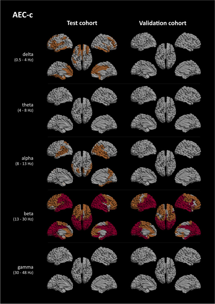
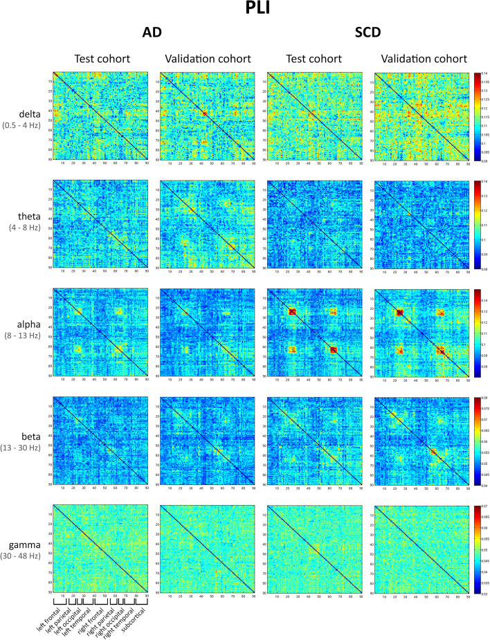
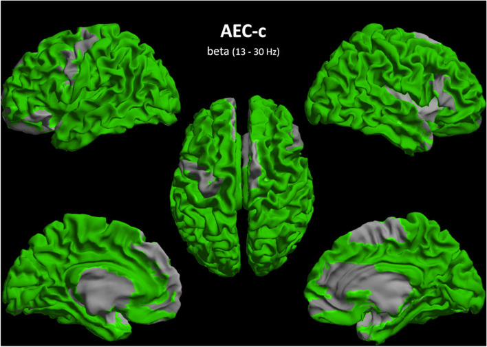
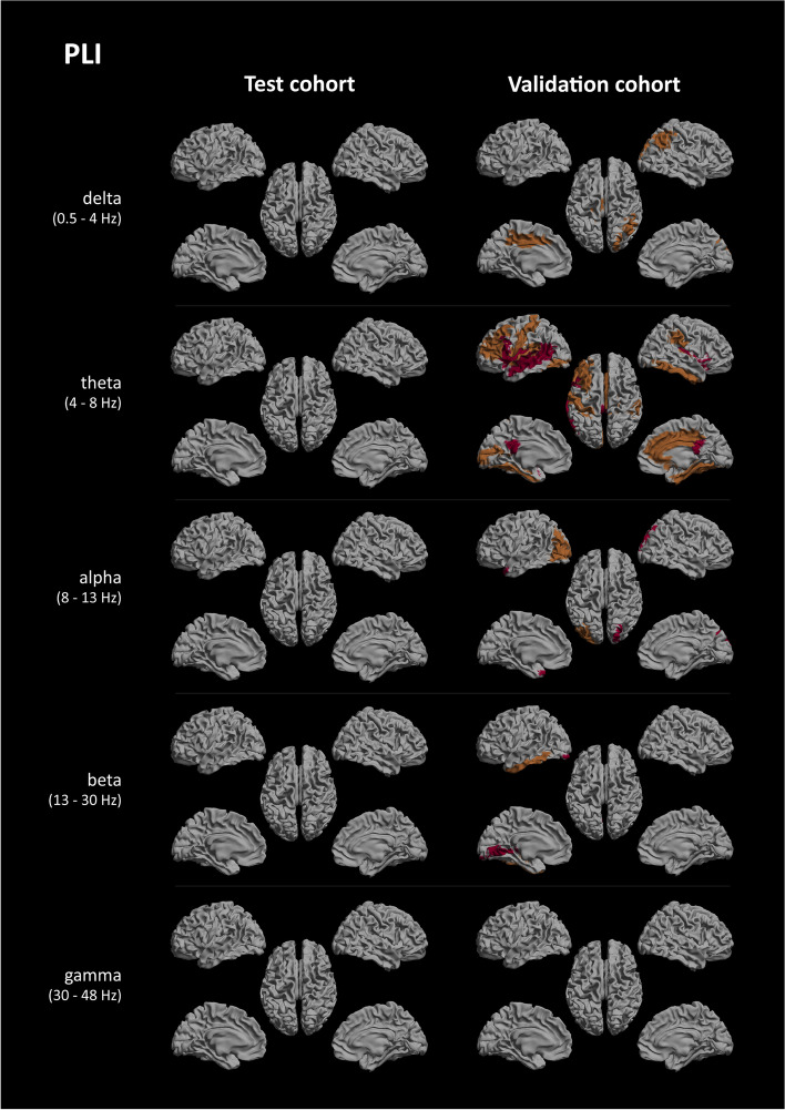
The AEC-c showed significantly and reproducibly lower global FC for the AD group compared to SCD, in the alpha (8-13 Hz) and beta (13-30 Hz) bands, while the PLI revealed reproducibly lower FC for the AD group in the delta (0.5-4 Hz) band and higher FC for the theta (4-8 Hz) band. Regionally, the beta band AEC-c showed reproducibility for almost all ROIs (except for 13 ROIs in the frontal and temporal lobes). For the other bands, the AEC-c and PLI did not show regional reproducibility after FDR correction. The theta band PLI was susceptible to the effect of relative power.
For MEG, the AEC-c is a sensitive and reproducible metric, able to distinguish FC differences between patients with AD dementia and cognitively healthy controls. These two measures likely reflect different aspects of neural activity and show differential sensitivity to changes in neural dynamics.
在阿尔茨海默病(AD)中,对功能性脑网络的分析受到缺乏可重复但有效的功能连接(FC)度量的限制。本研究旨在评估校正后的幅度包络相关(AEC-c)和相位滞后指数(PLI)这两种不受容积传导和场扩散影响的 FC 度量在两个分别患有 AD 痴呆和健康老年人对照的队列中的敏感性和可重复性。
经生物标志物证实的 AD 痴呆临床诊断患者和主观认知下降(SCD)对照组进行两次 5 分钟静息态 MEG 记录。数据由一个测试队列(AD=28;SCD=29)和一个验证队列(AD=29;SCD=27)组成。对自动解剖标记(AAL)图谱中的 90 个感兴趣区(ROI)进行时间序列估计。对于五个典型频带中的每一个,在所有 90 个 ROI 之间计算 AEC-c 和 PLI,并对每个 ROI 进行平均。构建广义线性模型以比较组间的全局 FC 差异,评估可重复性,并评估年龄和相对功率的影响。使用 Mann-Whitney U 检验评估区域 FC 差异的可重复性,并使用错误发现率(FDR)进行多重检验校正。
与 SCD 相比,AD 组的 AEC-c 在 alpha(8-13 Hz)和 beta(13-30 Hz)频段表现出显著且可重复的低全局 FC,而 PLI 则在 delta(0.5-4 Hz)频段表现出 AD 组可重复的低 FC 和 theta(4-8 Hz)频段的高 FC。在区域上,beta 频段的 AEC-c 对几乎所有 ROI 都具有可重复性(额叶和颞叶的 13 个 ROI 除外)。对于其他频段,在 FDR 校正后,AEC-c 和 PLI 均未显示区域可重复性。theta 频段的 PLI 易受相对功率的影响。
对于 MEG,AEC-c 是一种敏感且可重复的度量标准,能够区分 AD 痴呆患者和认知健康对照者之间的 FC 差异。这两种测量方法可能反映了神经活动的不同方面,并对神经动力学的变化表现出不同的敏感性。