Division of Infection and Immunity, The Roslin Institute & R(D)SVS, University of Edinburgh, Easter Bush, Midlothian, EH25 9RG, UK.
Novozymes A/S, Animal Health and Nutrition, 2800, Lyngby, Denmark.
Vet Res. 2022 Mar 2;53(1):15. doi: 10.1186/s13567-022-01033-0.
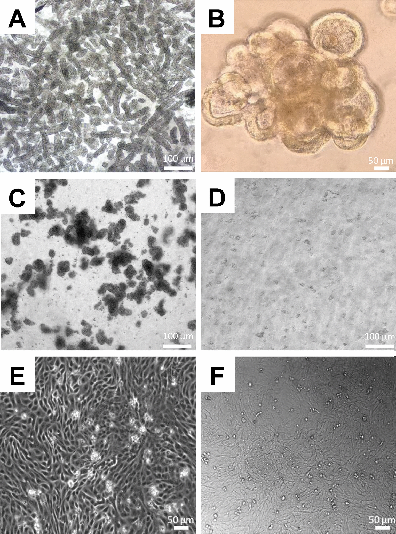
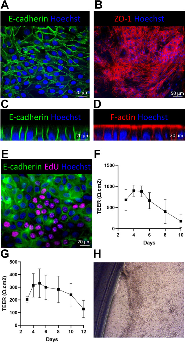
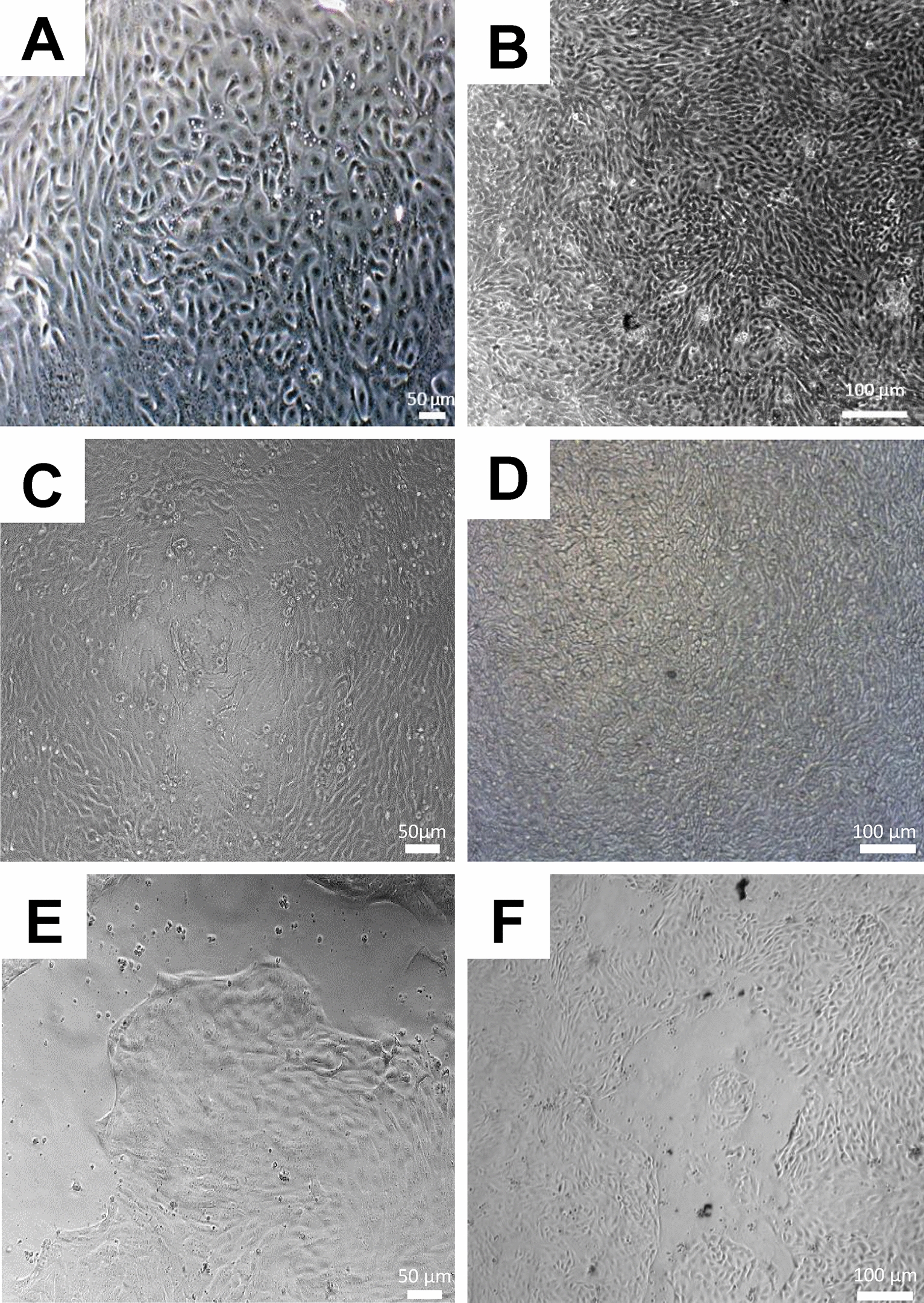
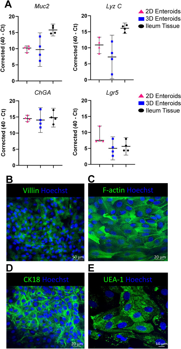
Three-dimensional (3D) intestinal enteroids are powerful in vitro models for studying intestinal biology. However, due to their closed structure direct access to the apical surface is impeded, limiting high-throughput applications of exogenous compounds and pathogens. In this study, we describe a method for generating confluent 2D enteroids from single-cell suspensions of enzymatically-dissociated ileum-derived bovine 3D enteroids. Confluent monolayers were first achieved using IntestiCult media but to establish a defined, cost-effective culture media, we also developed a bovine enteroid monolayer (BEM) medium. The monolayers cultured in BEM media proliferated extensively and formed confluent cell layers on both Matrigel-coated plastic plates and transwell inserts by day 3 of culture. The 2D enteroids maintained the epithelial cell lineages found in 3D enteroids and ileum tissue. In addition, the monolayers formed a functional epithelial barrier based on the presence of the adherens and tight junction proteins, E-cadherin and ZO-1, and electrical resistance across the monolayer was measured from day 3 and maintained for up to 7 days in culture. The method described here will provide a useful model to study bovine epithelial cell biology with ease of access to the apical surface of epithelial cells and has potential to investigate host-pathogen interactions and screen bioactive compounds.
三维(3D)肠类器官是研究肠道生物学的强大体外模型。然而,由于其封闭的结构,直接接触顶膜受到阻碍,限制了外源性化合物和病原体的高通量应用。在这项研究中,我们描述了一种从酶解的牛 3D 肠类器官的单细胞悬浮液中生成单层 2D 肠类器官的方法。首先使用 IntestiCult 培养基实现了单层细胞的融合,但为了建立一个明确的、具有成本效益的培养基,我们还开发了一种牛肠类器官单层(BEM)培养基。在 BEM 培养基中培养的单层细胞大量增殖,并在培养第 3 天在 Matrigel 包被的塑料板和 Transwell 插入物上形成融合的细胞层。2D 肠类器官保持了在 3D 肠类器官和回肠组织中发现的上皮细胞谱系。此外,单层细胞基于黏着连接和紧密连接蛋白 E-钙黏蛋白和 ZO-1 的存在形成了功能性的上皮屏障,并且从第 3 天开始测量单层的跨膜电阻,并在培养中维持长达 7 天。这里描述的方法将提供一个有用的模型,以方便地研究牛的上皮细胞生物学,并且具有研究宿主-病原体相互作用和筛选生物活性化合物的潜力。