Wang Tao, Xu Wei, Zhao Xintong, Bai Baoshuai, Hua Yuejie, Tang Jincheng, Chen Feifan, Liu Yu, Wang Yahui, Zhou Guangdong, Cao Yilin
Research Institute of Plastic Surgery, Wei Fang Medical University, Wei Fang, Shandong, 261041, PR China.
National Tissue Engineering Center of China, Shanghai, 200241, PR China.
Mater Today Bio. 2022 Mar 8;14:100234. doi: 10.1016/j.mtbio.2022.100234. eCollection 2022 Mar.
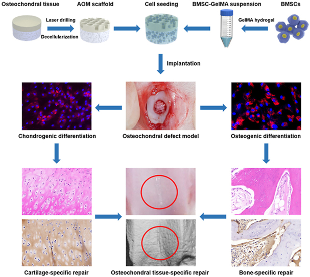
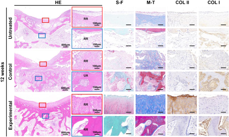
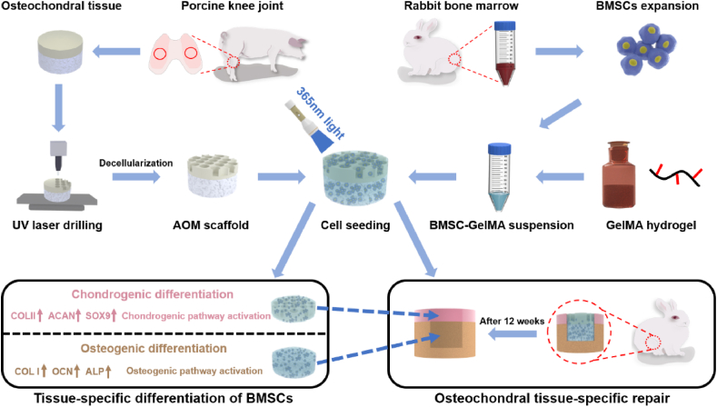
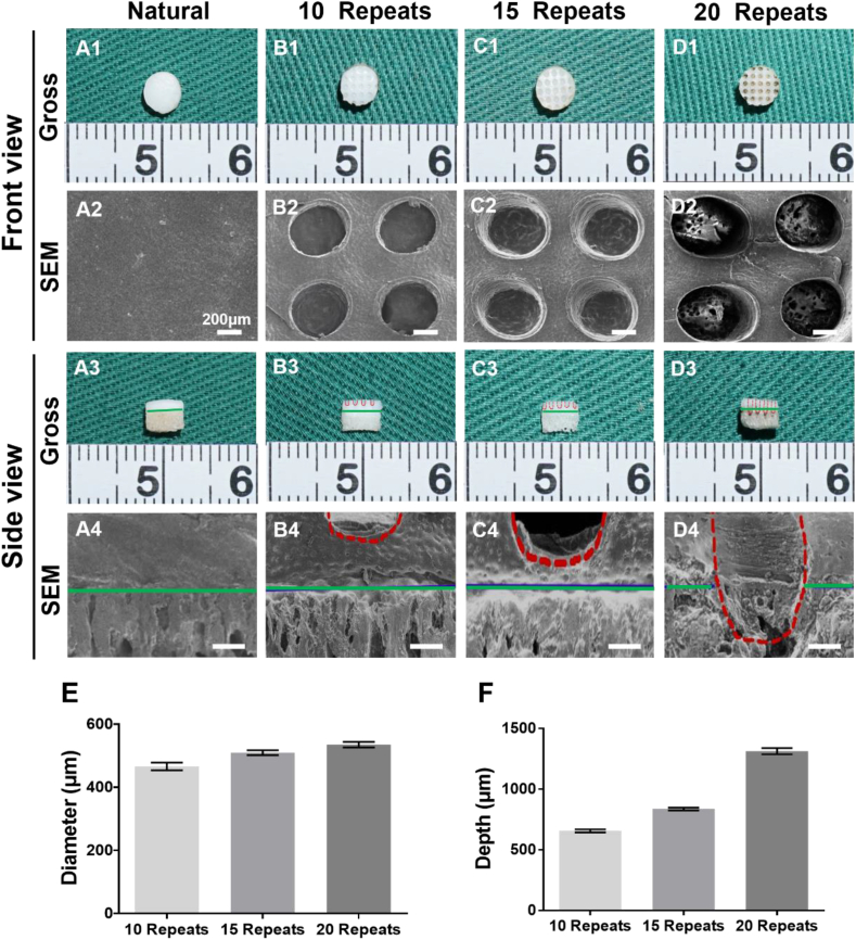
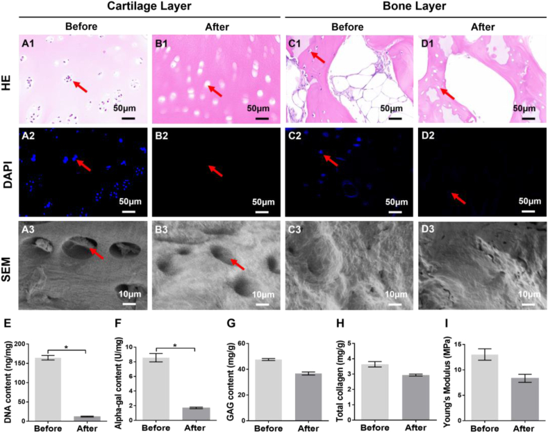
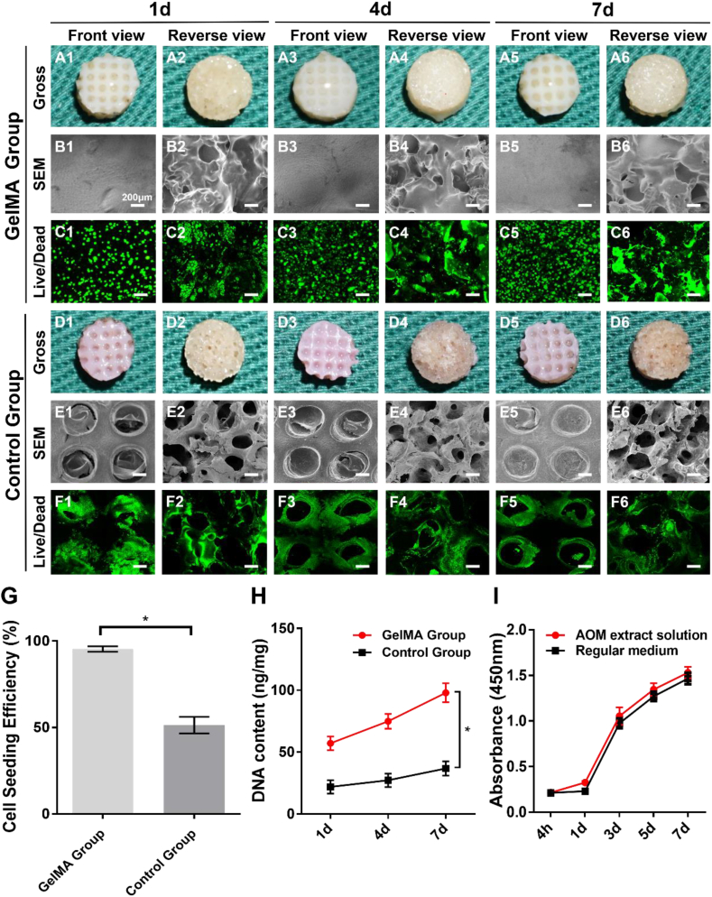
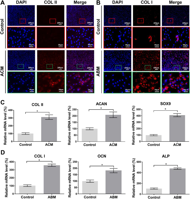
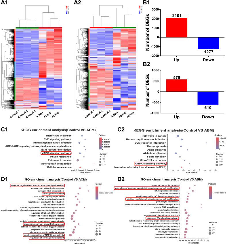
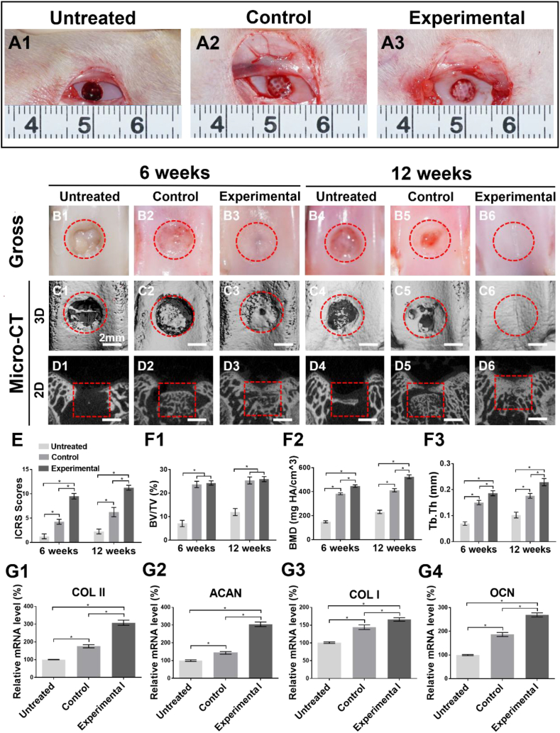
Tissue engineering provides a new approach for the treatment of osteochondral defects. However, the lack of an ideal double-layer scaffold with osteochondral-biomimetic microenvironment and interface similar to native articular tissue greatly limits clinical translation. Our current study developed a double-layer acellular osteochondral matrix (AOM) scaffold with natural osteochondral-biomimetic microenvironment and interface by integrating ultraviolet (UV) laser and decellularization techniques. The laser parameters were optimized to achieve a proper pore depth close to the osteochondral interface, which guaranteed complete decellularization, sufficient space for cell loading, and relative independence of the chondrogenic and osteogenic microenvironments. Gelatin-methacryloyl (GelMA) hydrogel was further used as the cell carrier to significantly enhance the efficiency and homogeneity of cell loading in the AOM scaffold with large pore structure. Additionally, results demonstrated that the components of the AOM scaffold could efficiently regulate the chondrogenic/osteogenic differentiations of bone marrow stromal cells (BMSCs) by activating the chondrogenic/osteogenic related pathways. Importantly, the AOM scaffolds combined with BMSC-laden GelMA hydrogel successfully realized tissue-specific repair of the osteochondral defects in a knee joint model of rabbit. The current study developed a novel double-layer osteochondral biomimetic scaffold and feasible strategy, providing strong support for the tissue-specific repair of osteochondral defects and its future clinical translation.
组织工程为治疗骨软骨缺损提供了一种新方法。然而,缺乏具有与天然关节组织相似的骨软骨仿生微环境和界面的理想双层支架极大地限制了其临床转化。我们目前的研究通过整合紫外线(UV)激光和脱细胞技术,开发了一种具有天然骨软骨仿生微环境和界面的双层脱细胞骨软骨基质(AOM)支架。优化激光参数以实现接近骨软骨界面的合适孔深度,这保证了完全脱细胞、足够的细胞接种空间以及软骨生成和成骨微环境的相对独立性。明胶-甲基丙烯酰基(GelMA)水凝胶进一步用作细胞载体,以显著提高细胞接种到具有大孔结构的AOM支架中的效率和均匀性。此外,结果表明AOM支架的成分可通过激活软骨生成/成骨相关途径有效调节骨髓间充质干细胞(BMSC)的软骨生成/成骨分化。重要的是,AOM支架与负载BMSC的GelMA水凝胶相结合,在兔膝关节模型中成功实现了骨软骨缺损的组织特异性修复。本研究开发了一种新型双层骨软骨仿生支架和可行策略,为骨软骨缺损的组织特异性修复及其未来临床转化提供了有力支持。