Wu Zhenquan, Cai Wenjia, Xie Hai, Chen Shida, Wang Yanbing, Lei Baiying, Zheng Yingfeng, Lu Lin
State Key Laboratory of Ophthalmology, Zhongshan Ophthalmic Center, Sun Yat-sen University, Guangzhou, China.
Health Science Center, School of Biomedical Engineering, Shenzhen University, Shenzhen, China.
Front Med (Lausanne). 2022 Mar 3;9:842680. doi: 10.3389/fmed.2022.842680. eCollection 2022.
To develop an artificial intelligence (AI) system that can predict optical coherence tomography (OCT)-derived high myopia grades based on fundus photographs.
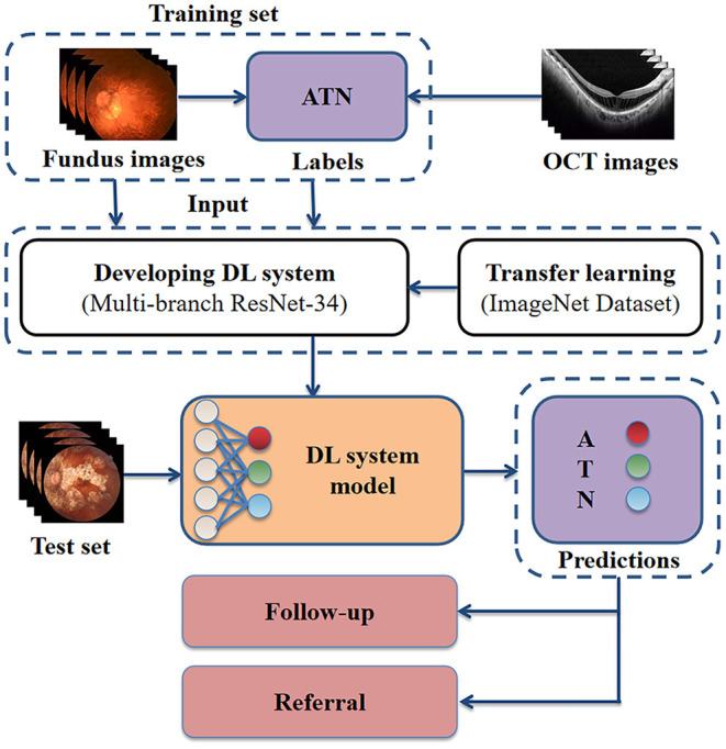
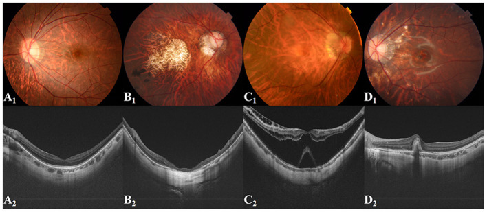
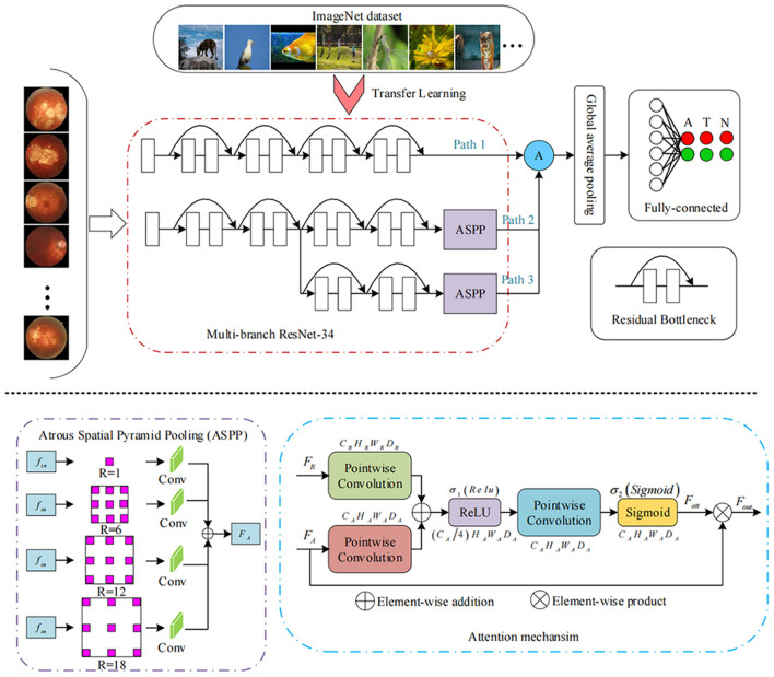
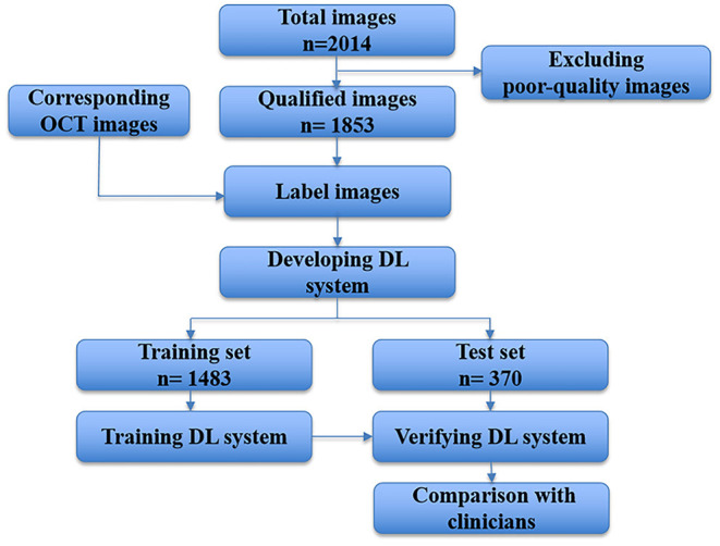
In this retrospective study, 1,853 qualified fundus photographs obtained from the Zhongshan Ophthalmic Center (ZOC) were selected to develop an AI system. Three retinal specialists assessed corresponding OCT images to label the fundus photographs. We developed a novel deep learning model to detect and predict myopic maculopathy according to the atrophy (A), traction (T), and neovascularisation (N) classification and grading system. Furthermore, we compared the performance of our model with that of ophthalmologists.
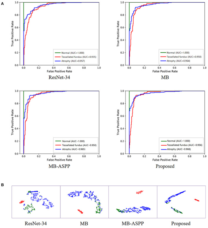
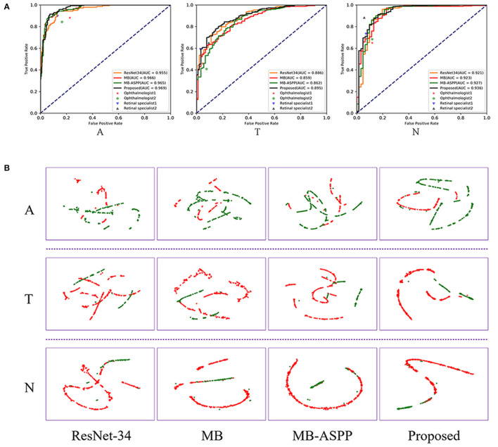
When evaluated on the test set, the deep learning model showed an area under the receiver operating characteristic curve (AUC) of 0.969 for category A, 0.895 for category T, and 0.936 for category N. The average accuracy of each category was 92.38% (A), 85.34% (T), and 94.21% (N). Moreover, the performance of our AI system was superior to that of attending ophthalmologists and comparable to that of retinal specialists.
Our AI system achieved performance comparable to that of retinal specialists in predicting vision-threatening conditions in high myopia via simple fundus photographs instead of fundus and OCT images. The application of this system can save the cost of patients' follow-up, and is more suitable for applications in less developed areas that only have fundus photography.
开发一种人工智能(AI)系统,该系统能够基于眼底照片预测光学相干断层扫描(OCT)得出的高度近视度数。
在这项回顾性研究中,从中山眼科中心(ZOC)选取了1853张合格的眼底照片来开发AI系统。三位视网膜专家评估相应的OCT图像以标记眼底照片。我们开发了一种新型深度学习模型,根据萎缩(A)、牵拉(T)和新生血管形成(N)分类及分级系统来检测和预测近视性黄斑病变。此外,我们将我们模型的性能与眼科医生的性能进行了比较。
在测试集上进行评估时,深度学习模型在A类中的受试者操作特征曲线下面积(AUC)为0.969,T类为0.895,N类为0.936。每类的平均准确率分别为92.38%(A)、85.34%(T)和94.21%(N)。此外,我们的AI系统的性能优于主治眼科医生,与视网膜专家相当。
我们的AI系统通过简单的眼底照片而非眼底和OCT图像,在预测高度近视中威胁视力的情况方面达到了与视网膜专家相当的性能。该系统的应用可以节省患者随访的成本,并且更适合在仅有眼底摄影的欠发达地区应用。