Graças Amanda M, Souza Willy P, Canut Ana Carolina A, Franciss Maurice Y, Zilberstein Bruno
Division of General Surgery, Beneficência Portuguesa of São Paulo Hospital, Gastromed-Zilberstein Institute, São Paulo, Brazil.
Division of Gastrointestinal Surgery, Beneficência Portuguesa of São Paulo Hospital, Gastromed - Zilberstein Institute and São Leopoldo Mandic School of Medicine, Campinas, Brazil.
Front Surg. 2022 Mar 7;9:792243. doi: 10.3389/fsurg.2022.792243. eCollection 2022.
The present study analyzes diagnostic and therapeutic surgical aspects of primary small bowel melanoma, describing the clinical case and reviewing the literature. Malignant melanomas represent 1-3% of all malignant tumors of the gastrointestinal tract and are therefore uncommon. Only a few cases of metastatic melanoma (1-5%) are diagnosed in the early stages, while still asymptomatic. They show up as imaging exam findings and have better chance of treatment. Most of the time, the diagnosis is late and made in the presence of complications. The final diagnosis frequently depends on the surgical intervention after a serious complication.
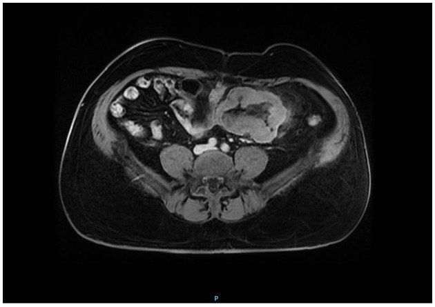
This case report refers to a 55-year-old male patient, complaining of abdominal pain, blackened stools, and palpable tumor in the abdomen for ~30 days. A tomography scan was performed, which revealed the thickening and parietal enhancement of the small intestine loops with mesenteric adenomegaly and intermingled collection. For diagnostic confirmation, a laparoscopy was performed, which revealed a tumor at the jejunal level, and its resection was performed in the same act. The anatomopathological examination revealed that it was a primary small bowel melanoma.
The bibliographic research of the small bowel melanoma demonstrated that, in this situation, the lesion can be interpreted as a primary site or metastatic lesion, considering the possibility of a single primary lesion, whose diagnosis becomes more laborious. In such cases, adjuvant therapy must be considered. The expected 5-year survival is about 9-13%.
本研究分析原发性小肠黑色素瘤的诊断和治疗手术方面,描述临床病例并复习文献。恶性黑色素瘤占胃肠道所有恶性肿瘤的1% - 3%,因此并不常见。只有少数转移性黑色素瘤病例(1% - 5%)在早期仍无症状时被诊断出来。它们表现为影像学检查结果,治疗机会更大。大多数情况下,诊断较晚,是在出现并发症时做出的。最终诊断往往取决于严重并发症后的手术干预。
本病例报告涉及一名55岁男性患者,主诉腹痛、黑便和腹部可触及肿物约30天。进行了断层扫描,显示小肠肠袢增厚和肠壁强化,伴有肠系膜淋巴结肿大和混杂积液。为确诊,进行了腹腔镜检查,发现空肠水平有一个肿瘤,并在同一手术中进行了切除。解剖病理学检查显示这是原发性小肠黑色素瘤。
小肠黑色素瘤的文献研究表明,在这种情况下,考虑到单一原发性病变的可能性,该病变可被解释为原发部位或转移病变,其诊断变得更加费力。在这种情况下,必须考虑辅助治疗。预期5年生存率约为9% - 13%。