Department of Electroceramics, Instituto de Cerámica y Vidrio-CSIC, Kelsen 5, 28049 Madrid, Spain.
Department of Immunology, School of Medicine, Universidad Complutense de Madrid, 12 de Octubre Health Research Institute (imas12), 28040 Madrid, Spain.
Int J Mol Sci. 2022 Mar 21;23(6):3410. doi: 10.3390/ijms23063410.
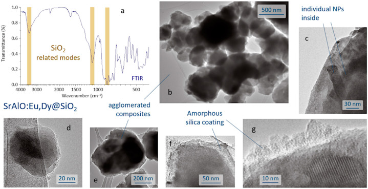
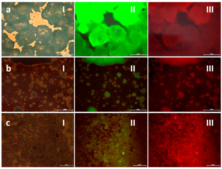
In recent decades, the demand for biomedical imaging tools has grown very rapidly as a key feature for biomedical research and diagnostic applications. Particularly, fluorescence imaging has gained increased attention as a non-invasive, inexpensive technique that allows real-time imaging. However, tissue auto-fluorescence under external illumination, together with a weak tissue penetration of low wavelength excitation light, largely restricts the application of the technique. Accordingly, new types of fluorescent labels are currently being investigated and, in this search, phosphorescent nanoparticles promise great potential, as they combine the interesting size-dependent properties of nanoscale materials with a long-lasting phosphorescence-type emission that allows optical imaging well after excitation (so avoiding autofluorescence). In this work, core-shell structures consisting of SrAlO:Eu,Dy luminescent cores encapsulated within a biocompatible silica shell were prepared, showing a green persistent phosphorescence with an afterglow time of more than 1000 s. A high-energy ball milling procedure was used to reduce the size of the starting phosphors to a size suitable for cellular uptake, while the silica coating was produced by a reverse micelle methodology that eventually allows the excitation and emission light to pass efficiently through the shell. Confocal fluorescence microscopy using HeLa cancer cells confirmed the potential of the all-ceramic composites produced as feasible labels for in vitro optical imaging.
近几十年来,生物医学成像工具的需求增长非常迅速,成为生物医学研究和诊断应用的关键特征。特别是荧光成像是一种非侵入性、低成本的技术,允许实时成像,因此受到了越来越多的关注。然而,组织在外部照明下的自发荧光,以及低波长激发光的弱组织穿透,在很大程度上限制了该技术的应用。因此,目前正在研究新型荧光标记物,在这一探索中,磷光纳米颗粒具有很大的潜力,因为它们将纳米材料的有趣的尺寸依赖性性质与持久的磷光型发射结合在一起,在激发后允许光学成像持续很长时间(从而避免自发荧光)。在这项工作中,制备了由 SrAlO:Eu,Dy 发光核包封在生物相容性硅壳内的核壳结构,表现出绿色持久磷光,余辉时间超过 1000 秒。采用高能球磨法将起始磷光体的尺寸减小到适合细胞摄取的尺寸,而硅壳则通过反胶束方法制备,最终允许激发和发射光有效地通过壳层。使用 HeLa 癌细胞的共焦荧光显微镜证实了所制备的全陶瓷复合材料作为体外光学成像可行标记物的潜力。