Wang Zhufeng, Chen Bigui, Fu Yu, Ou Changxing, Rong Qiuping, Kong Xuetao, Xu Wei, Deng Yangqing, Jiang Mei, Xie Jiaxing
National Clinical Research Center for Respiratory Disease, State Key Laboratory of Respiratory Disease, Guangzhou Institute of Respiratory Health, The First Affiliated Hospital of Guangzhou Medical University, Guangzhou, China.
Department of Respiratory and Critical Care Medicine, Yangjiang People's Hospital, Yangjiang, China.
Front Med (Lausanne). 2022 Mar 9;9:830754. doi: 10.3389/fmed.2022.830754. eCollection 2022.
Growing evidence added to the results from observational studies of lung cancer patients exhibiting eosinophilia. However, whether eosinophils contributed to tumor immune surveillance or neoplastic evolution was unknown. This study aimed to analyze the causal association between eosinophilia and lung cancer.
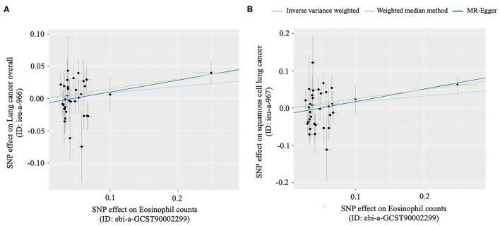
The causal effect of eosinophil count on lung cancer from a genome-wide association study (GWAS) was investigated using the two-sample Mendelian randomization (MR) method. Secondary results according to different histological subtypes of lung cancer were also implemented. Meanwhile, we compared the measured levels of blood eosinophil counts among different subtypes of lung cancer from real-world data.
The median absolute eosinophilic count (unit: 10/L) [median (min, max): Lung adenocarcinoma 0.7 (0.5, 15); Squamous cell lung cancer 0.7 (0.5, 1.3); Small cell lung cancer 0.7 (0.6, 1.3); = 0.96] and the median eosinophil to leukocyte ratio [median (min, max): Lung adenocarcinoma 8.7% (2.1, 42.2%); Squamous cell lung cancer 9.3% (4.1, 17.7%); Small cell lung cancer 8.9% (5.1, 24.1%); = 0.91] were similar among different histological subtypes of lung cancer. MR methods indicated that eosinophilia may provide 28% higher risk for squamous cell lung cancer in East Asian [Weighted median method: odds ratio (OR) = 1.28, 95% CI: 1.04-1.57, = 0.02].
Our study suggested that eosinophilia may be a potential causal risk factor in the progression of squamous cell lung cancer in East Asian.
越来越多的证据补充了对表现出嗜酸性粒细胞增多的肺癌患者的观察性研究结果。然而,嗜酸性粒细胞是否有助于肿瘤免疫监视或肿瘤演变尚不清楚。本研究旨在分析嗜酸性粒细胞增多与肺癌之间的因果关系。
使用两样本孟德尔随机化(MR)方法研究全基因组关联研究(GWAS)中嗜酸性粒细胞计数对肺癌的因果效应。还根据肺癌的不同组织学亚型得出了次要结果。同时,我们从实际数据中比较了不同亚型肺癌患者血液嗜酸性粒细胞计数的测量水平。
不同组织学亚型肺癌患者的嗜酸性粒细胞绝对计数中位数(单位:10⁹/L)[中位数(最小值,最大值):肺腺癌0.7(0.5,15);肺鳞状细胞癌0.7(0.5,1.3);小细胞肺癌0.7(0.6,1.3);P = 0.96]和嗜酸性粒细胞与白细胞比值中位数[中位数(最小值,最大值):肺腺癌8.7%(2.1,42.2%);肺鳞状细胞癌9.3%(4.1,17.7%);小细胞肺癌8.9%(5.1,24.‘1%);P = ’0.91]相似。MR方法表明,在东亚人群中,嗜酸性粒细胞增多可能使肺鳞状细胞癌风险增加28%[加权中位数法:优势比(OR)= 1.28,95%置信区间(CI):1.04 - 1.57,P = 0.02]。
我们的研究表明,嗜酸性粒细胞增多可能是东亚人群肺鳞状细胞癌进展的潜在因果危险因素。