MRC Laboratory of Molecular Biology, Francis Crick Avenue, Cambridge CB2 0QH, UK.
Ernst Ruska-Centrum für Mikroskopie und Spektroskopie mit Elektronen, Forschungszentrum Jülich GmbH, 52425 Jülich, Germany.
Ultramicroscopy. 2022 Jul;237:113510. doi: 10.1016/j.ultramic.2022.113510. Epub 2022 Mar 19.
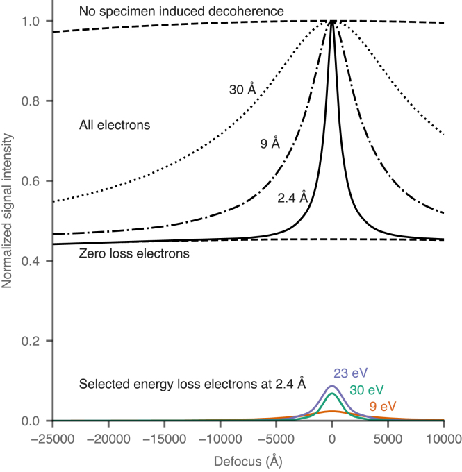
We investigate potential improvements in using electron cryomicroscopy to image thick specimens with high-resolution phase contrast imaging. In particular, using model experiments, electron scattering theory, Monte Carlo and multislice simulations, we determine the potential for improving electron cryomicrographs of proteins within a cell using chromatic aberration (C) correction. We show that inelastically scattered electrons lose a quantifiable amount of spatial coherence as they transit the specimen, yet can be used to enhance the signal from thick biological specimens (in the 1000 to 5000 Å range) provided they are imaged close to focus with an achromatic lens. This loss of information quantified here, which we call "specimen induced decoherence", is a fundamental limit on imaging biological molecules in situ. We further show that with foreseeable advances in transmission electron microscope technology, it should be possible to directly locate and uniquely identify sub-100 kDa proteins without the need for labels, in a vitrified specimen taken from a cell.
我们研究了在使用电子冷冻显微镜对厚标本进行高分辨率相衬成像时的潜在改进。特别是,我们使用模型实验、电子散射理论、蒙特卡罗和多层模拟,确定了使用色差(C)校正来改善细胞内蛋白质电子冷冻显微镜图像的潜力。我们表明,非弹性散射电子在穿过标本时会失去可量化的空间相干性,但如果使用消色差透镜在近焦处成像,它们可以用于增强厚生物标本(1000 至 5000 Å 范围内)的信号。我们在这里量化的这种信息损失,我们称之为“标本诱导去相干”,是对原位成像生物分子的基本限制。我们进一步表明,随着透射电子显微镜技术的可预见的进步,应该有可能在无需标记的情况下,直接定位和唯一识别小于 100 kDa 的蛋白质,而无需对取自细胞的玻璃化标本进行任何处理。