Naydenova Katerina, Kamegawa Akiko, Peet Mathew J, Henderson Richard, Fujiyoshi Yoshinori, Russo Christopher J
MRC Laboratory of Molecular Biology, Francis Crick Avenue, Cambridge CB2 0QH, UK.
Cellular and Structural Physiology Laboratory (CeSPL), Tokyo Medical and Dental University, Yushima, Bunkyo-ku, Tokyo, Japan.
Ultramicroscopy. 2022 Jul;237:113512. doi: 10.1016/j.ultramic.2022.113512. Epub 2022 Mar 19.
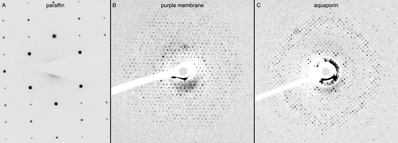
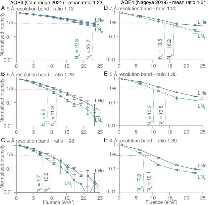
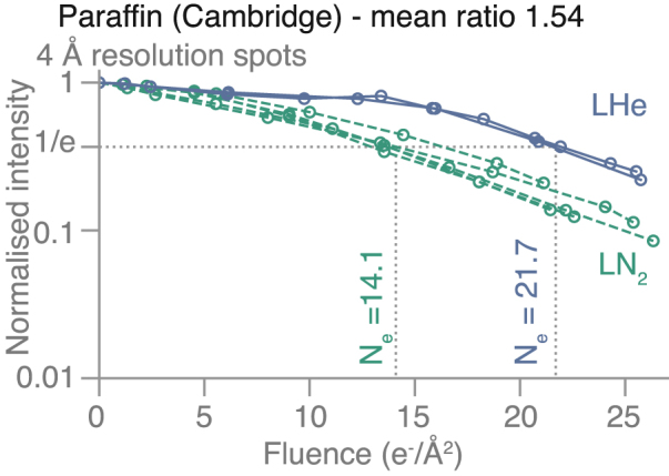
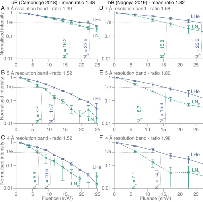
We have studied the fading of electron diffraction spots from two-dimensional (2D) crystals of paraffin (CH), purple membrane (bacteriorhodopsin) and aquaporin 4 (AQP4) at stage temperatures between 4K and 100K. We observed that the diffraction spots at resolutions between 3 Å and 20 Å fade more slowly at liquid-helium temperatures compared to liquid-nitrogen temperatures, by a factor of between 1.2 and 1.8, depending on the specimens. If the reduction in the effective rate of radiation damage for 2D crystals at liquid-helium temperature (as measured by spot fading) can be shown to extend to macromolecular assemblies embedded in amorphous ice, this would suggest that valuable improvements to electron cryomicroscopy (cryoEM) of biological specimens could be made by reducing the temperature of the specimens under irradiation below what is obtainable using standard liquid-nitrogen cryostats.
我们研究了石蜡(CH)、紫膜(细菌视紫红质)和水通道蛋白4(AQP4)二维(2D)晶体的电子衍射斑点在4K至100K的阶段温度下的衰减情况。我们观察到,在3埃至20埃分辨率下的衍射斑点,在液氦温度下比液氮温度下褪色更慢,根据样本不同,慢1.2至1.8倍。如果能证明在液氦温度下二维晶体辐射损伤有效率的降低(通过斑点褪色测量)可扩展到嵌入非晶冰中的大分子聚集体,这将表明通过将辐照下样本的温度降低到使用标准液氮低温恒温器所能达到的温度以下,生物样本的电子冷冻显微镜(cryoEM)可能会有显著改进。