• 文献检索
  • 文档翻译
  • 深度研究
  • 学术资讯
  • Suppr Zotero 插件Zotero 插件
  • 邀请有礼
  • 套餐&价格
  • 历史记录
应用&插件
Suppr Zotero 插件Zotero 插件浏览器插件Mac 客户端Windows 客户端微信小程序
定价
高级版会员购买积分包购买API积分包
服务
文献检索文档翻译深度研究API 文档MCP 服务
关于我们
关于 Suppr公司介绍联系我们用户协议隐私条款
关注我们

Suppr 超能文献

核心技术专利:CN118964589B侵权必究
粤ICP备2023148730 号-1Suppr @ 2026

文献检索

告别复杂PubMed语法,用中文像聊天一样搜索,搜遍4000万医学文献。AI智能推荐,让科研检索更轻松。

立即免费搜索

文件翻译

保留排版,准确专业,支持PDF/Word/PPT等文件格式,支持 12+语言互译。

免费翻译文档

深度研究

AI帮你快速写综述,25分钟生成高质量综述,智能提取关键信息,辅助科研写作。

立即免费体验

水凝胶在基于支架的生物打印中的关键作用综合评估

A Comprehensive Assessment on the Pivotal Role of Hydrogels in Scaffold-Based Bioprinting.

作者信息

Parimala Chelvi Ratnamani Matangi, Zhang Xinping, Wang Hongjun

机构信息

Department of Biomedical Engineering, Stevens Institute of Technology, Hoboken, NJ 07030, USA.

Department of Orthopaedics, Center for Musculoskeletal Research, University of Rochester Medical Center, Rochester, NY 14642, USA.

出版信息

Gels. 2022 Apr 13;8(4):239. doi: 10.3390/gels8040239.

DOI:10.3390/gels8040239
PMID:35448140
原文链接:https://pmc.ncbi.nlm.nih.gov/articles/PMC9028353/
Abstract

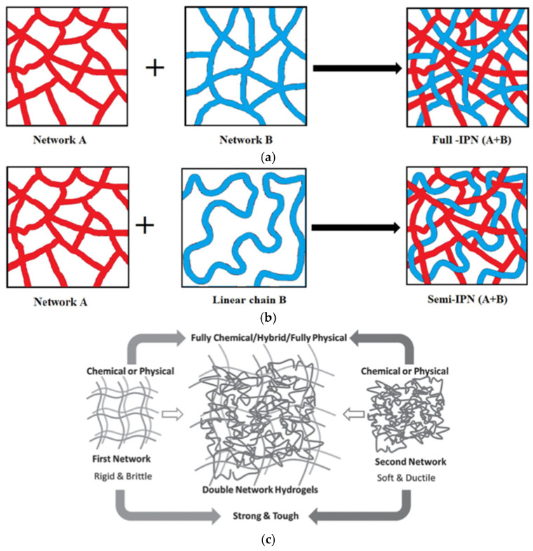
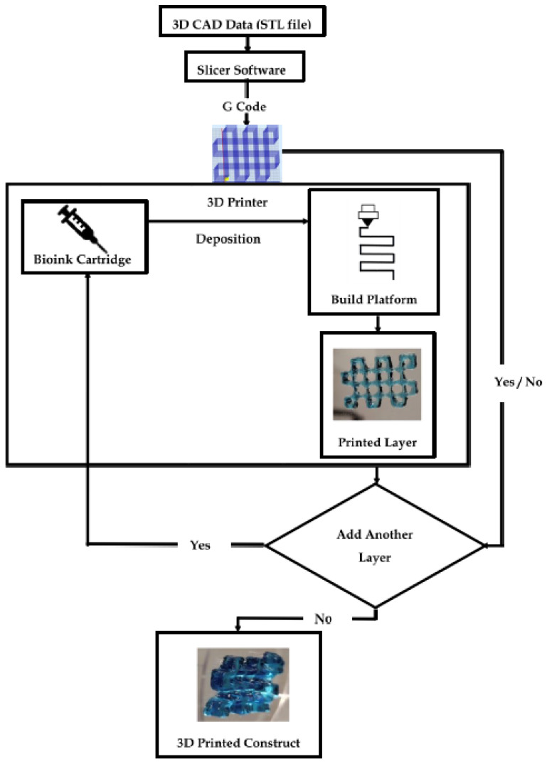
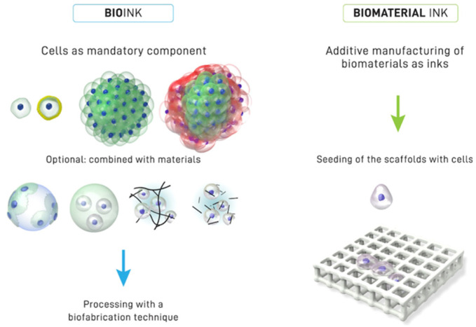
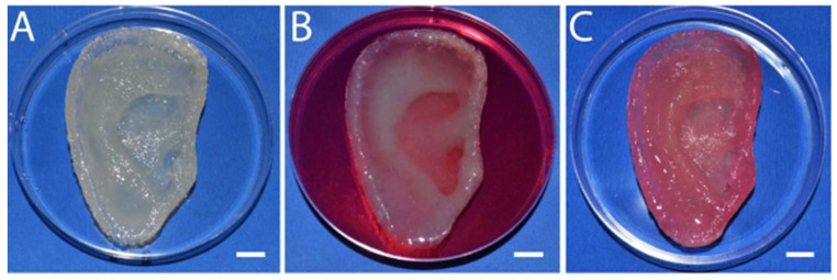
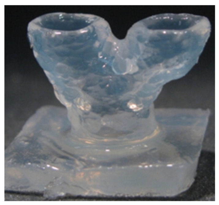
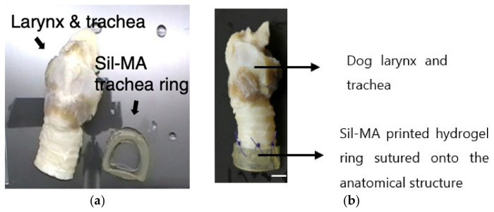
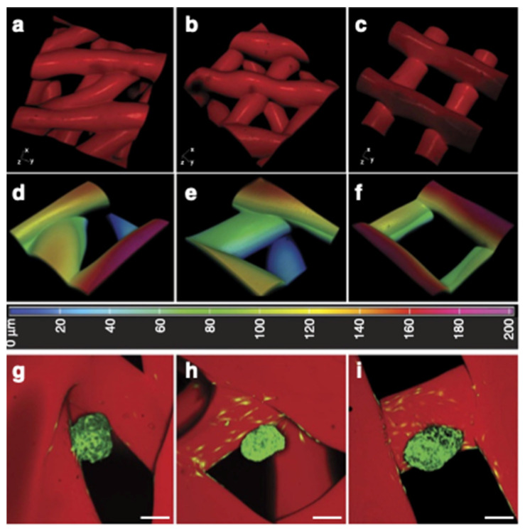
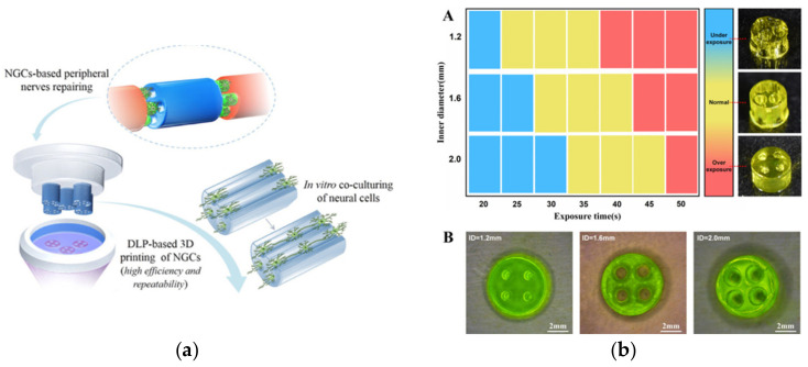
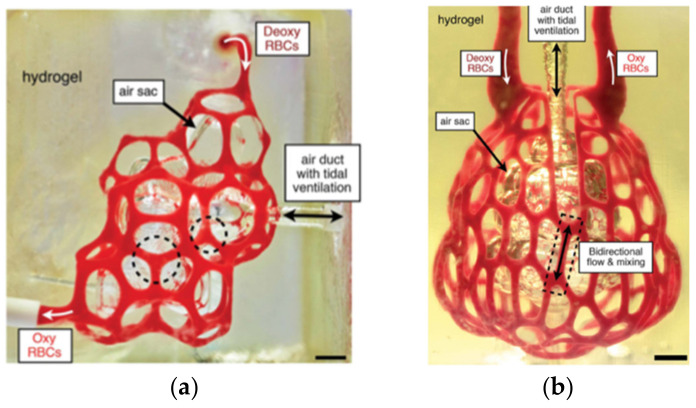
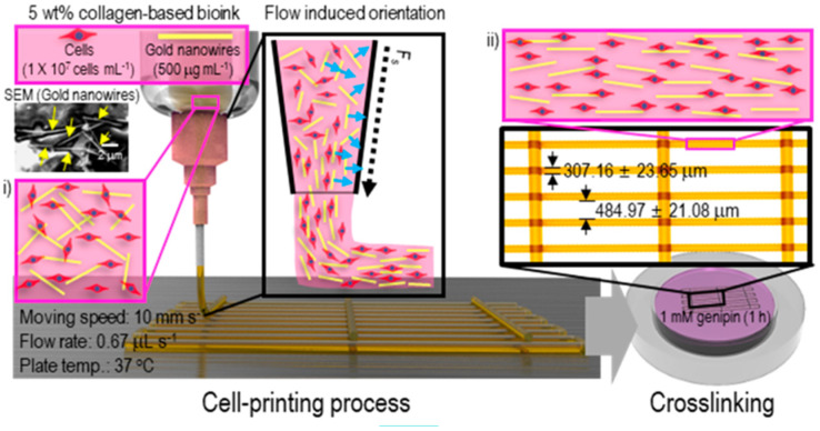
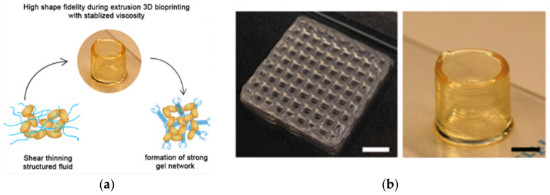
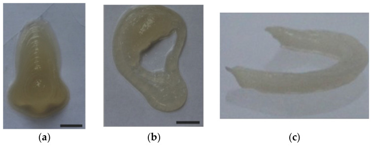
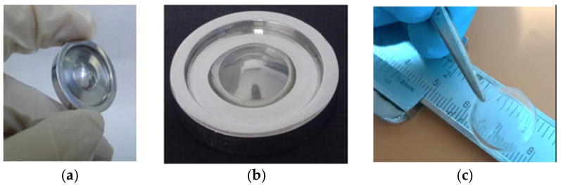
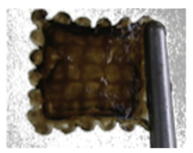
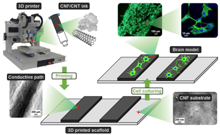
The past a few decades have seen exponential growth in the field of regenerative medicine. What began as extirpative (complete tissue or organ removal), with little regard to the effects of tissue loss and/or disfigurement, has evolved towards fabricating engineered tissues using personalized living cells (e.g., stem cells), and customizing a matrix or structural organization to support and guide tissue development. Biofabrication, largely accomplished through three-dimensional (3D) printing technology, provides precise, controlled, and layered assemblies of cells and biomaterials, emulating the heterogenous microenvironment of the in vivo tissue architecture. This review provides a concise framework for the bio-manufacturing process and addresses the contributions of hydrogels to biological modeling. The versatility of hydrogels in bioprinting is detailed along with an extensive elaboration of their physical, mechanical, and biological properties, as well as their assets and limitations in bioprinting. The scope of various hydrogels in tissue formation has been discussed through the case studies of biofabricated 3D constructs in order to provide the readers with a glimpse into the barrier-breaking accomplishments of biomedical sciences. In the end, the restraints of bioprinting itself are discussed, accompanied with the identification of available engineering strategies to overcome them.

摘要

在过去几十年里,再生医学领域呈指数级增长。最初的治疗方式是切除(完全去除组织或器官),几乎不考虑组织损失和/或毁容的影响,如今已发展为利用个性化活细胞(如干细胞)制造工程组织,并定制基质或结构组织以支持和引导组织发育。生物制造主要通过三维(3D)打印技术完成,可提供精确、可控且分层的细胞和生物材料组装体,模拟体内组织结构的异质微环境。本综述为生物制造过程提供了一个简明框架,并探讨了水凝胶对生物建模的贡献。详细介绍了水凝胶在生物打印中的多功能性,以及其物理、机械和生物学特性,及其在生物打印中的优势和局限性。通过生物制造3D构建体的案例研究,讨论了各种水凝胶在组织形成中的应用范围,以便让读者了解生物医学科学的突破性成就。最后,讨论了生物打印本身的限制,并确定了可用的工程策略来克服这些限制。

https://cdn.ncbi.nlm.nih.gov/pmc/blobs/ccf5/9028353/d8a3df6f55d7/gels-08-00239-g022.jpg
https://cdn.ncbi.nlm.nih.gov/pmc/blobs/ccf5/9028353/55fcfa810598/gels-08-00239-g020.jpg
https://cdn.ncbi.nlm.nih.gov/pmc/blobs/ccf5/9028353/acf73a7e543c/gels-08-00239-g001.jpg
https://cdn.ncbi.nlm.nih.gov/pmc/blobs/ccf5/9028353/b09bed42b7c6/gels-08-00239-g002.jpg
https://cdn.ncbi.nlm.nih.gov/pmc/blobs/ccf5/9028353/eeeb665800da/gels-08-00239-g003.jpg
https://cdn.ncbi.nlm.nih.gov/pmc/blobs/ccf5/9028353/824a49a220ee/gels-08-00239-g004.jpg
https://cdn.ncbi.nlm.nih.gov/pmc/blobs/ccf5/9028353/a58d871721fb/gels-08-00239-g005.jpg
https://cdn.ncbi.nlm.nih.gov/pmc/blobs/ccf5/9028353/b1d140364e42/gels-08-00239-g006.jpg
https://cdn.ncbi.nlm.nih.gov/pmc/blobs/ccf5/9028353/ffa1e45e3a2c/gels-08-00239-g007.jpg
https://cdn.ncbi.nlm.nih.gov/pmc/blobs/ccf5/9028353/ddd2220eb2fb/gels-08-00239-g008.jpg
https://cdn.ncbi.nlm.nih.gov/pmc/blobs/ccf5/9028353/c4ed48419128/gels-08-00239-g009.jpg
https://cdn.ncbi.nlm.nih.gov/pmc/blobs/ccf5/9028353/5d3cb43c3b9e/gels-08-00239-g010.jpg
https://cdn.ncbi.nlm.nih.gov/pmc/blobs/ccf5/9028353/6efc8a2e1803/gels-08-00239-g011.jpg
https://cdn.ncbi.nlm.nih.gov/pmc/blobs/ccf5/9028353/7e4bf273b479/gels-08-00239-g012.jpg
https://cdn.ncbi.nlm.nih.gov/pmc/blobs/ccf5/9028353/c6a9aedc49f6/gels-08-00239-g013.jpg
https://cdn.ncbi.nlm.nih.gov/pmc/blobs/ccf5/9028353/ce15f909a482/gels-08-00239-g014.jpg
https://cdn.ncbi.nlm.nih.gov/pmc/blobs/ccf5/9028353/4f3fc57652ef/gels-08-00239-g015.jpg
https://cdn.ncbi.nlm.nih.gov/pmc/blobs/ccf5/9028353/c207ba9bf7e8/gels-08-00239-g016.jpg
https://cdn.ncbi.nlm.nih.gov/pmc/blobs/ccf5/9028353/36bd3c2e304d/gels-08-00239-g017.jpg
https://cdn.ncbi.nlm.nih.gov/pmc/blobs/ccf5/9028353/7fd7c3dd867f/gels-08-00239-g018.jpg
https://cdn.ncbi.nlm.nih.gov/pmc/blobs/ccf5/9028353/929acba4e12c/gels-08-00239-g019.jpg
https://cdn.ncbi.nlm.nih.gov/pmc/blobs/ccf5/9028353/b334bf160003/gels-08-00239-g021.jpg
https://cdn.ncbi.nlm.nih.gov/pmc/blobs/ccf5/9028353/d8a3df6f55d7/gels-08-00239-g022.jpg

相似文献

1
A Comprehensive Assessment on the Pivotal Role of Hydrogels in Scaffold-Based Bioprinting.水凝胶在基于支架的生物打印中的关键作用综合评估
Gels. 2022 Apr 13;8(4):239. doi: 10.3390/gels8040239.
2
Embedded bioprinting for designer 3D tissue constructs with complex structural organization.嵌入式生物打印用于具有复杂结构组织的设计 3D 组织构建体。
Acta Biomater. 2022 Mar 1;140:1-22. doi: 10.1016/j.actbio.2021.11.048. Epub 2021 Dec 5.
3
Advancing bioinks for 3D bioprinting using reactive fillers: A review.使用反应性填料推进用于3D生物打印的生物墨水:综述。
Acta Biomater. 2020 Sep 1;113:1-22. doi: 10.1016/j.actbio.2020.06.040. Epub 2020 Jul 2.
4
Converging functionality: Strategies for 3D hybrid-construct biofabrication and the role of composite biomaterials for skeletal regeneration.汇聚功能:用于 3D 混合结构生物制造的策略和用于骨骼再生的复合生物材料的作用。
Acta Biomater. 2021 Sep 15;132:188-216. doi: 10.1016/j.actbio.2021.03.008. Epub 2021 Mar 10.
5
Bioprintable tough hydrogels for tissue engineering applications.用于组织工程应用的可生物打印坚韧水凝胶。
Adv Colloid Interface Sci. 2020 Jul;281:102163. doi: 10.1016/j.cis.2020.102163. Epub 2020 Apr 25.
6
Modeling and Fabrication of Silk Fibroin-Gelatin-Based Constructs Using Extrusion-Based Three-Dimensional Bioprinting.基于挤出式三维生物打印技术的丝素蛋白-明胶基构建体的建模与制造
ACS Biomater Sci Eng. 2021 Jul 12;7(7):3306-3320. doi: 10.1021/acsbiomaterials.1c00410. Epub 2021 Jun 8.
7
3D bioprinting of complex channels within cell-laden hydrogels.细胞负载水凝胶内复杂通道的三维生物打印。
Acta Biomater. 2019 Sep 1;95:214-224. doi: 10.1016/j.actbio.2019.02.038. Epub 2019 Mar 1.
8
Progress in 3D bioprinting technology for tissue/organ regenerative engineering.用于组织/器官再生工程的3D生物打印技术进展。
Biomaterials. 2020 Jan;226:119536. doi: 10.1016/j.biomaterials.2019.119536. Epub 2019 Oct 11.
9
3D Bioprinting of Human Tissues: Biofabrication, Bioinks, and Bioreactors.三维生物打印人体组织:生物制造、生物墨水和生物反应器。
Int J Mol Sci. 2021 Apr 12;22(8):3971. doi: 10.3390/ijms22083971.
10
3D bioprinting of molecularly engineered PEG-based hydrogels utilizing gelatin fragments.利用明胶片段对基于聚乙二醇的分子工程水凝胶进行 3D 生物打印。
Biofabrication. 2021 Aug 5;13(4). doi: 10.1088/1758-5090/ac0ff0.

引用本文的文献

1
Hydrogel-Based Scaffolds: Advancing Bone Regeneration Through Tissue Engineering.基于水凝胶的支架:通过组织工程促进骨再生
Gels. 2025 Feb 27;11(3):175. doi: 10.3390/gels11030175.
2
Advances in 3D printing combined with tissue engineering for nerve regeneration and repair.3D打印与组织工程相结合在神经再生与修复方面的进展。
J Nanobiotechnology. 2025 Jan 3;23(1):5. doi: 10.1186/s12951-024-03052-9.
3
Exploring Synergistic Effects of Bioprinted Extracellular Vesicles for Skin Regeneration.探索生物打印细胞外囊泡对皮肤再生的协同作用。

本文引用的文献

1
Nanocomposite Bioinks Based on Agarose and 2D Nanosilicates with Tunable Flow Properties and Bioactivity for 3D Bioprinting.基于琼脂糖和二维纳米硅酸盐的具有可调流动特性和生物活性的纳米复合生物墨水用于3D生物打印
ACS Appl Bio Mater. 2019 Feb 18;2(2):796-806. doi: 10.1021/acsabm.8b00665. Epub 2019 Jan 24.
2
Natural Biomaterials and Their Use as Bioinks for Printing Tissues.天然生物材料及其作为组织打印生物墨水的用途。
Bioengineering (Basel). 2021 Feb 20;8(2):27. doi: 10.3390/bioengineering8020027.
3
Expansion of Ovarian Cancer Stem-like Cells in Poly(ethylene glycol)-Cross-Linked Poly(methyl vinyl ether--maleic acid) and Alginate Double-Network Hydrogels.
Biomedicines. 2024 Jul 18;12(7):1605. doi: 10.3390/biomedicines12071605.
4
Poly(Vinyl Alcohol)/Bovine Serum Albumin Hybrid Hydrogels with Tunable Mechanical Properties.具有可调机械性能的聚乙烯醇/牛血清白蛋白杂化水凝胶
Polymers (Basel). 2023 Dec 4;15(23):4611. doi: 10.3390/polym15234611.
5
Artificial Intelligence-Empowered 3D and 4D Printing Technologies toward Smarter Biomedical Materials and Approaches.人工智能赋能的3D和4D打印技术助力更智能的生物医学材料与方法
Polymers (Basel). 2022 Jul 8;14(14):2794. doi: 10.3390/polym14142794.
6
A Guide to Polysaccharide-Based Hydrogel Bioinks for 3D Bioprinting Applications.多糖基水凝胶生物墨水用于 3D 生物打印应用指南。
Int J Mol Sci. 2022 Jun 12;23(12):6564. doi: 10.3390/ijms23126564.
聚(乙二醇)交联聚(甲基乙烯基醚-马来酸)与海藻酸盐双网络水凝胶中卵巢癌干细胞样细胞的扩增
ACS Biomater Sci Eng. 2020 Jun 8;6(6):3310-3326. doi: 10.1021/acsbiomaterials.9b01967. Epub 2020 Apr 21.
4
3D Bioprinting of Spatially Heterogeneous Collagen Constructs for Cartilage Tissue Engineering.用于软骨组织工程的空间异质胶原蛋白构建体的3D生物打印
ACS Biomater Sci Eng. 2016 Oct 10;2(10):1800-1805. doi: 10.1021/acsbiomaterials.6b00288. Epub 2016 Aug 4.
5
Processing and Properties of Chitosan Inks for 3D Printing of Hydrogel Microstructures.用于水凝胶微结构3D打印的壳聚糖墨水的加工与性能
ACS Biomater Sci Eng. 2018 Jul 9;4(7):2643-2652. doi: 10.1021/acsbiomaterials.8b00415. Epub 2018 Jun 7.
6
Cellular extrusion bioprinting improves kidney organoid reproducibility and conformation.细胞挤出式生物打印提高了肾类器官的重现性和形态。
Nat Mater. 2021 Feb;20(2):260-271. doi: 10.1038/s41563-020-00853-9. Epub 2020 Nov 23.
7
Perfluorocarbon-based oxygen carriers: from physics to physiology.基于全氟碳的氧载体:从物理学到生理学。
Pflugers Arch. 2021 Feb;473(2):139-150. doi: 10.1007/s00424-020-02482-2. Epub 2020 Nov 3.
8
Silk fibroin nanofibers: a promising ink additive for extrusion three-dimensional bioprinting.丝素蛋白纳米纤维:一种用于挤出式三维生物打印的有前景的墨水添加剂。
Mater Today Bio. 2020 Sep 19;8:100078. doi: 10.1016/j.mtbio.2020.100078. eCollection 2020 Sep.
9
Extrusion 3D Printing of Polymeric Materials with Advanced Properties.具有先进性能的聚合物材料的挤出3D打印
Adv Sci (Weinh). 2020 Aug 5;7(17):2001379. doi: 10.1002/advs.202001379. eCollection 2020 Sep.
10
3D Printed Silicone Meniscus Implants: Influence of the 3D Printing Process on Properties of Silicone Implants.3D打印硅胶半月板植入物:3D打印工艺对硅胶植入物性能的影响。
Polymers (Basel). 2020 Sep 18;12(9):2136. doi: 10.3390/polym12092136.