Peng Wei, Hao Qinghong, Gao Heng, Wang Yang, Wang Jun, Tu Yang, Yu Siyi, Li Hui, Zhu Tianmin
School of Acupuncture and Tuina, Chengdu University of Traditional Chinese Medicine, Chengdu, China.
School of Rehabilitation and Health Preservation, Chengdu University of Traditional Chinese Medicine, Chengdu, China.
Front Neurol. 2022 Apr 18;13:841514. doi: 10.3389/fneur.2022.841514. eCollection 2022.
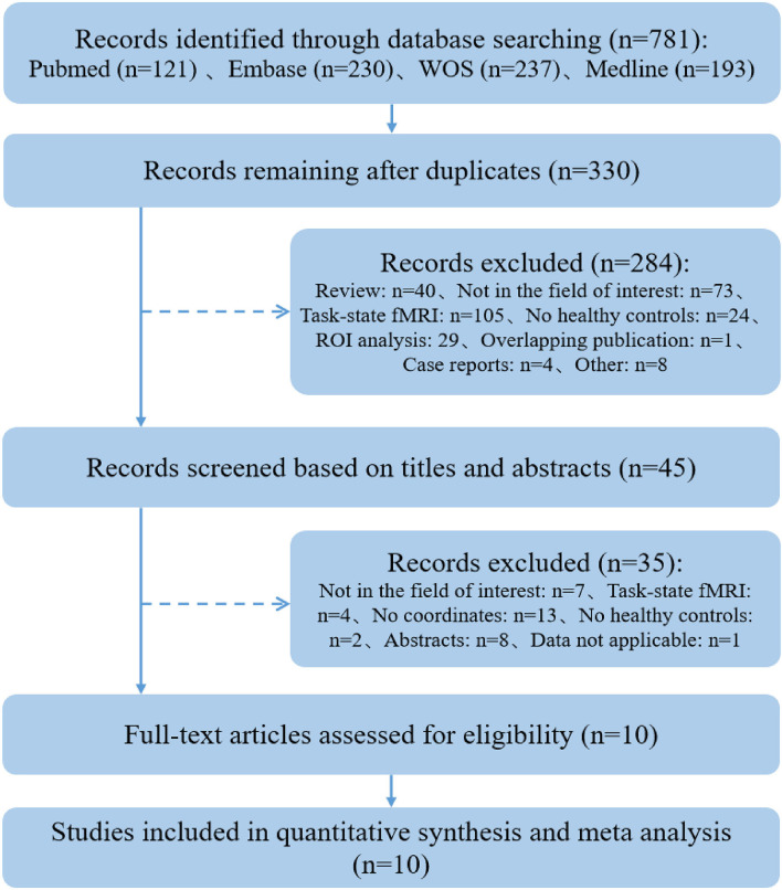
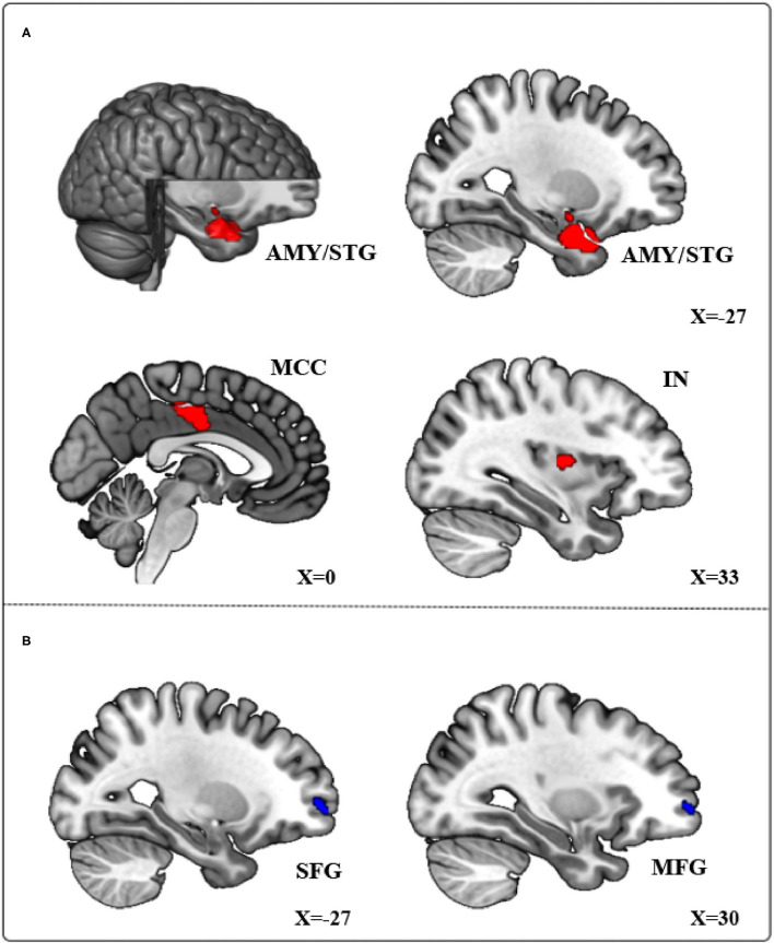
Previous resting-state functional MRI (fMRI) studies found spontaneous neural activity in the brains of Pathological Internet Use (PIU) subjects. However, the findings were inconsistent in studies using different neuroimaging analyses. This meta-analytic study aimed to identify a common pattern of altered brain activity from different studies. Resting-state fMRI studies, based on whole-brain analysis methods published before July 1, 2021, were searched in multiple databases (PubMed, EMBASE, MEDLINE, and Web of Science). A voxel-based signed differential mapping (SDM) method was used to clarify brain regions, which showed anomalous activity in PIU subjects compared with healthy controls (HCs). Ten eligible publications consisting of 306 PIU subjects and 314 HCs were included in the SDM meta-analysis. Compared with HCs, subjects with PIU showed increased spontaneous neural functional activity in the left temporal pole of the superior temporal cortex, left amygdala, bilateral median cingulate cortex, and right insula. Meanwhile, a decreased spontaneous neural activity was identified in the left dorsolateral superior frontal gyrus and right middle frontal gyrus in the subjects with PIU. These abnormal brain regions are associated with cognitive executive control and emotional regulation. The consistent changes under different functional brain imaging indicators found in our study may provide important targets for the future diagnosis and intervention of PIU. www.crd.york.ac.uk/PROSPERO, identifier: CRD42021258119.
先前的静息态功能磁共振成像(fMRI)研究发现,病理性互联网使用(PIU)受试者大脑中存在自发神经活动。然而,在使用不同神经影像学分析方法的研究中,结果并不一致。这项荟萃分析研究旨在从不同研究中确定大脑活动改变的共同模式。在多个数据库(PubMed、EMBASE、MEDLINE和Web of Science)中检索了基于2021年7月1日前发表的全脑分析方法的静息态fMRI研究。采用基于体素的符号差异映射(SDM)方法来明确与健康对照(HC)相比,PIU受试者中显示异常活动的脑区。SDM荟萃分析纳入了10篇符合条件的出版物,包括306名PIU受试者和314名HC。与HC相比,PIU受试者在颞上叶皮质的左侧颞极、左侧杏仁核、双侧扣带回中部皮质和右侧岛叶表现出自发神经功能活动增加。同时,在PIU受试者的左侧背外侧额上回和右侧额中回发现自发神经活动减少。这些异常脑区与认知执行控制和情绪调节有关。我们的研究中在不同功能性脑成像指标下发现的一致变化可能为未来PIU的诊断和干预提供重要靶点。www.crd.york.ac.uk/PROSPERO,标识符:CRD42021258119。