• 文献检索
  • 文档翻译
  • 深度研究
  • 学术资讯
  • Suppr Zotero 插件Zotero 插件
  • 邀请有礼
  • 套餐&价格
  • 历史记录
应用&插件
Suppr Zotero 插件Zotero 插件浏览器插件Mac 客户端Windows 客户端微信小程序
定价
高级版会员购买积分包购买API积分包
服务
文献检索文档翻译深度研究API 文档MCP 服务
关于我们
关于 Suppr公司介绍联系我们用户协议隐私条款
关注我们

Suppr 超能文献

核心技术专利:CN118964589B侵权必究
粤ICP备2023148730 号-1Suppr @ 2026

文献检索

告别复杂PubMed语法,用中文像聊天一样搜索,搜遍4000万医学文献。AI智能推荐,让科研检索更轻松。

立即免费搜索

文件翻译

保留排版,准确专业,支持PDF/Word/PPT等文件格式,支持 12+语言互译。

免费翻译文档

深度研究

AI帮你快速写综述,25分钟生成高质量综述,智能提取关键信息,辅助科研写作。

立即免费体验

单细胞分析人类硬脑膜和脑膜瘤揭示细胞脑膜景观,并深入了解脑膜瘤免疫反应。

Single-cell profiling of human dura and meningioma reveals cellular meningeal landscape and insights into meningioma immune response.

机构信息

Department of Neurological Surgery, Washington University School of Medicine, St. Louis, MO, USA.

Department of Pathology and Immunology, Washington University School of Medicine, St. Louis, MO, USA.

出版信息

Genome Med. 2022 May 10;14(1):49. doi: 10.1186/s13073-022-01051-9.

DOI:10.1186/s13073-022-01051-9
PMID:35534852
原文链接:https://pmc.ncbi.nlm.nih.gov/articles/PMC9088131/
Abstract

BACKGROUND

Recent investigations of the meninges have highlighted the importance of the dura layer in central nervous system immune surveillance beyond a purely structural role. However, our understanding of the meninges largely stems from the use of pre-clinical models rather than human samples.

METHODS

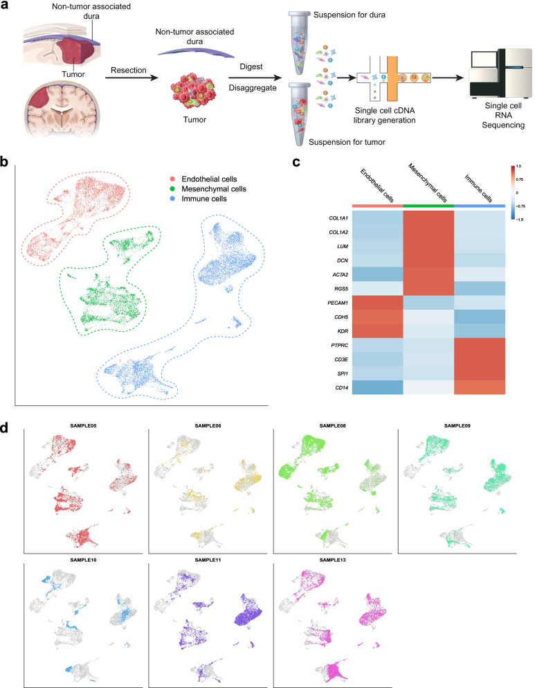
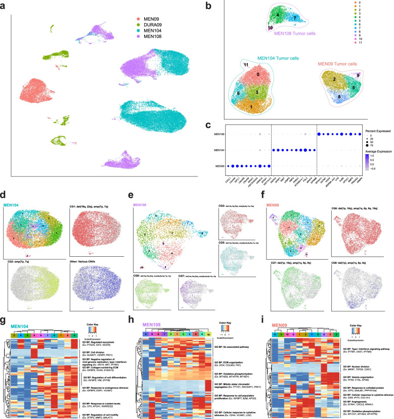
Single-cell RNA sequencing of seven non-tumor-associated human dura samples and six primary meningioma tumor samples (4 matched and 2 non-matched) was performed. Cell type identities, gene expression profiles, and T cell receptor expression were analyzed. Copy number variant (CNV) analysis was performed to identify putative tumor cells and analyze intratumoral CNV heterogeneity. Immunohistochemistry and imaging mass cytometry was performed on selected samples to validate protein expression and reveal spatial localization of select protein markers.

RESULTS

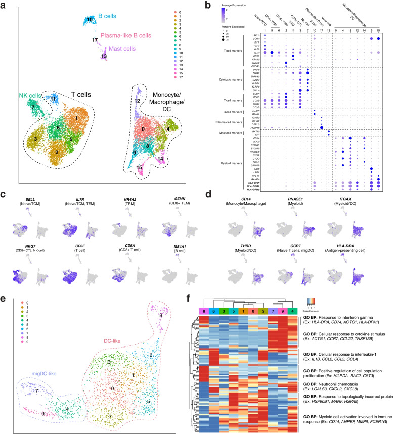
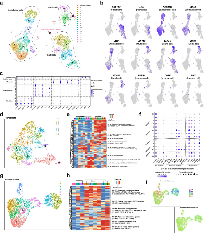
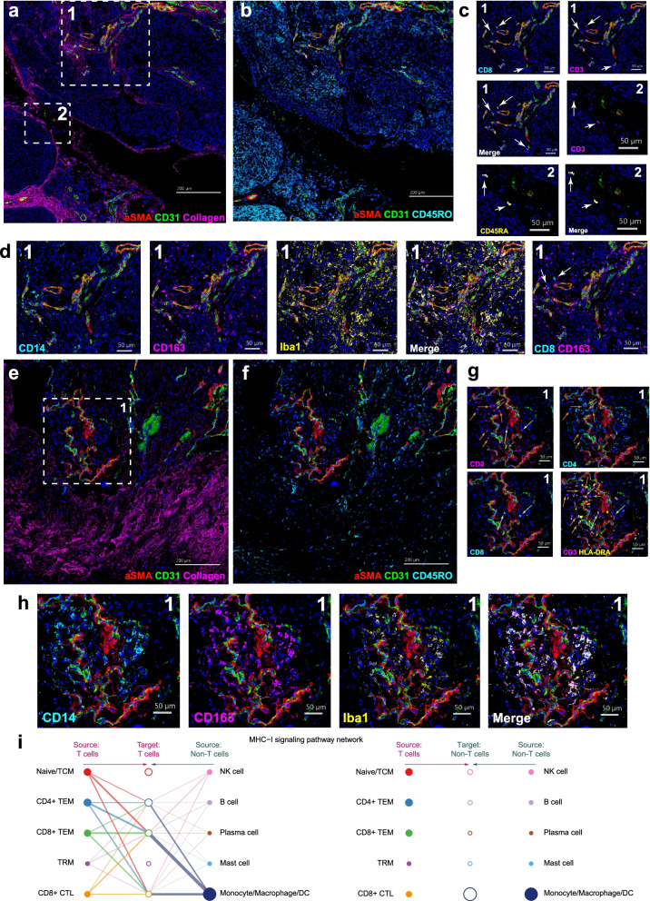
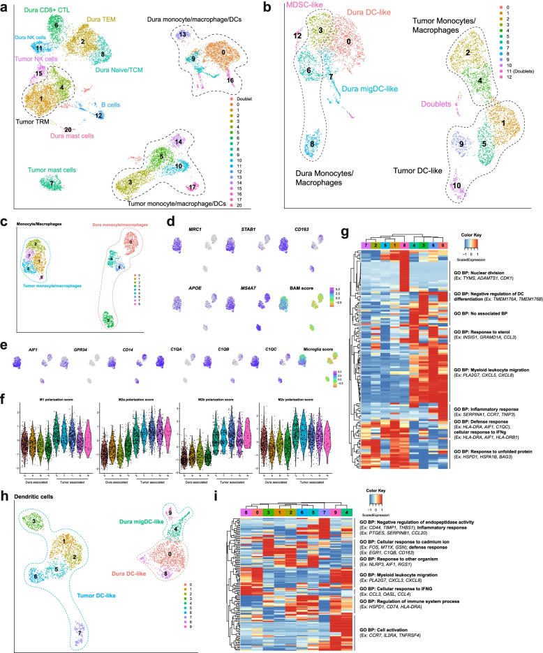
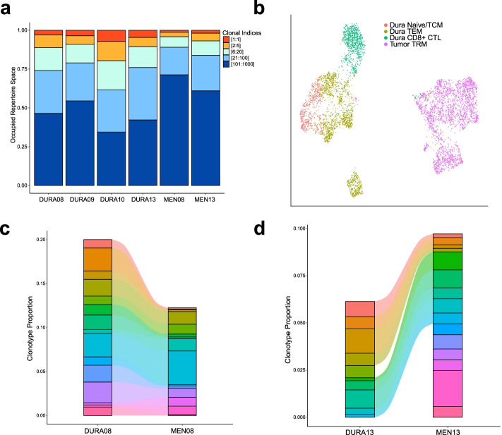
In this study, we use single-cell RNA sequencing to perform the first characterization of both non-tumor-associated human dura and primary meningioma samples. First, we reveal a complex immune microenvironment in human dura that is transcriptionally distinct from that of meningioma. In addition, we characterize a functionally diverse and heterogenous landscape of non-immune cells including endothelial cells and fibroblasts. Through imaging mass cytometry, we highlight the spatial relationship among immune cell types and vasculature in non-tumor-associated dura. Utilizing T cell receptor sequencing, we show significant TCR overlap between matched dura and meningioma samples. Finally, we report copy number variant heterogeneity within our meningioma samples.

CONCLUSIONS

Our comprehensive investigation of both the immune and non-immune cellular landscapes of human dura and meningioma at single-cell resolution builds upon previously published data in murine models and provides new insight into previously uncharacterized roles of human dura.

摘要

背景

最近对脑膜的研究强调了硬脑膜层在中枢神经系统免疫监视中的重要性,超出了纯粹的结构作用。然而,我们对脑膜的理解主要来自于临床前模型的应用,而不是人类样本。

方法

对 7 个非肿瘤相关的人类硬脑膜样本和 6 个原发性脑膜瘤肿瘤样本(4 个匹配和 2 个非匹配)进行单细胞 RNA 测序。分析细胞类型身份、基因表达谱和 T 细胞受体表达。进行拷贝数变异(CNV)分析,以识别潜在的肿瘤细胞并分析肿瘤内 CNV 异质性。对选定的样本进行免疫组织化学和成像质谱细胞术检测,以验证蛋白质表达并揭示选择蛋白标记物的空间定位。

结果

在这项研究中,我们使用单细胞 RNA 测序首次对非肿瘤相关的人类硬脑膜和原发性脑膜瘤样本进行了特征描述。首先,我们揭示了人类硬脑膜中复杂的免疫微环境,其转录组与脑膜瘤明显不同。此外,我们还描述了一种功能多样且异质的非免疫细胞景观,包括内皮细胞和成纤维细胞。通过成像质谱细胞术,我们突出了非肿瘤相关硬脑膜中免疫细胞类型与脉管系统之间的空间关系。利用 T 细胞受体测序,我们显示了匹配的硬脑膜和脑膜瘤样本之间存在显著的 TCR 重叠。最后,我们报告了我们脑膜瘤样本中的拷贝数变异异质性。

结论

我们对人类硬脑膜和脑膜瘤的免疫和非免疫细胞景观进行了单细胞分辨率的全面研究,这是对以前在鼠模型中发表的数据的扩展,并为以前未被描述的人类硬脑膜的作用提供了新的见解。

https://cdn.ncbi.nlm.nih.gov/pmc/blobs/76ad/9088131/85f1e61b6e07/13073_2022_1051_Fig7_HTML.jpg
https://cdn.ncbi.nlm.nih.gov/pmc/blobs/76ad/9088131/20fe31007f14/13073_2022_1051_Fig1_HTML.jpg
https://cdn.ncbi.nlm.nih.gov/pmc/blobs/76ad/9088131/b33283510a06/13073_2022_1051_Fig2_HTML.jpg
https://cdn.ncbi.nlm.nih.gov/pmc/blobs/76ad/9088131/b56f36cf6fae/13073_2022_1051_Fig3_HTML.jpg
https://cdn.ncbi.nlm.nih.gov/pmc/blobs/76ad/9088131/0d751ea2f99c/13073_2022_1051_Fig4_HTML.jpg
https://cdn.ncbi.nlm.nih.gov/pmc/blobs/76ad/9088131/18c784e0b37f/13073_2022_1051_Fig5_HTML.jpg
https://cdn.ncbi.nlm.nih.gov/pmc/blobs/76ad/9088131/b9d3c13042ee/13073_2022_1051_Fig6_HTML.jpg
https://cdn.ncbi.nlm.nih.gov/pmc/blobs/76ad/9088131/85f1e61b6e07/13073_2022_1051_Fig7_HTML.jpg

相似文献

1
Single-cell profiling of human dura and meningioma reveals cellular meningeal landscape and insights into meningioma immune response.单细胞分析人类硬脑膜和脑膜瘤揭示细胞脑膜景观,并深入了解脑膜瘤免疫反应。
Genome Med. 2022 May 10;14(1):49. doi: 10.1186/s13073-022-01051-9.
2
DNA methylation profile of human dura and leptomeninges.人脑硬脑膜和软脑膜的 DNA 甲基化图谱。
J Neuropathol Exp Neurol. 2023 Jun 20;82(7):641-649. doi: 10.1093/jnen/nlad036.
3
Single-cell RNA sequencing identifies macrophage signatures correlated with clinical features and tumour microenvironment in meningiomas.单细胞 RNA 测序鉴定出与脑膜瘤临床特征和肿瘤微环境相关的巨噬细胞特征。
IET Syst Biol. 2023 Oct;17(5):259-270. doi: 10.1049/syb2.12074. Epub 2023 Jul 29.
4
Decoding meningioma heterogeneity and neoplastic cell-macrophage interaction through single-cell transcriptome profiling across pathological grades.通过对不同病理分级的单细胞转录组谱分析,解析脑膜瘤异质性和肿瘤细胞-巨噬细胞相互作用。
J Transl Med. 2023 Oct 25;21(1):751. doi: 10.1186/s12967-023-04445-4.
5
Leveraging single-cell sequencing to classify and characterize tumor subgroups in bulk RNA-sequencing data.利用单细胞测序对批量 RNA 测序数据中的肿瘤亚群进行分类和特征分析。
J Neurooncol. 2024 Jul;168(3):515-524. doi: 10.1007/s11060-024-04710-6. Epub 2024 May 29.
6
Associations of pathological diagnosis and genetic abnormalities in meningiomas with the embryological origins of the meninges.脑膜瘤的病理诊断和遗传异常与脑膜的胚胎起源的关系。
Sci Rep. 2021 Mar 26;11(1):6987. doi: 10.1038/s41598-021-86298-9.
7
Prominent meningeal enhancement adjacent to meningioma on Gd-DTPA-enhanced MR images: histopathologic correlation.钆喷酸葡胺增强磁共振成像(Gd-DTPA-enhanced MR images)上脑膜瘤附近明显的脑膜强化:组织病理学相关性
Radiology. 1990 May;175(2):431-3. doi: 10.1148/radiology.175.2.2326470.
8
Histone deacetylase inhibitor AR-42 differentially affects cell-cycle transit in meningeal and meningioma cells, potently inhibiting NF2-deficient meningioma growth.组蛋白去乙酰化酶抑制剂 AR-42 可差异化影响脑膜和脑膜瘤细胞的细胞周期进程,强力抑制 NF2 缺失型脑膜瘤的生长。
Cancer Res. 2013 Jan 15;73(2):792-803. doi: 10.1158/0008-5472.CAN-12-1888. Epub 2012 Nov 14.
9
First insight into the somatic mutation burden of neurofibromatosis type 2-associated grade I and grade II meningiomas: a case report comprehensive genomic study of two cranial meningiomas with vastly different clinical presentation.对2型神经纤维瘤病相关的I级和II级脑膜瘤体细胞突变负担的初步见解:一项对两例临床表现差异极大的颅脑膜瘤的病例报告及综合基因组研究。
BMC Cancer. 2017 Feb 13;17(1):127. doi: 10.1186/s12885-017-3127-6.
10
MRI of papillary meningiomas in children.儿童乳头状脑膜瘤的磁共振成像
Pediatr Radiol. 1995 Nov;25 Suppl 1:S9-13.

引用本文的文献

1
Immunological and pathological characteristics of brain parenchymal and leptomeningeal metastases from non-small cell lung cancer.非小细胞肺癌脑实质和软脑膜转移的免疫和病理特征
Cell Discov. 2025 Aug 29;11(1):72. doi: 10.1038/s41421-025-00828-7.
2
Spatially Encoded Oncogenesis and Transcriptional Plasticity in Meningioma: Drivers of Therapeutic Resistance and Opportunities for Targeted Intervention.脑膜瘤中的空间编码肿瘤发生与转录可塑性:治疗耐药的驱动因素及靶向干预机会
Cancers (Basel). 2025 Aug 19;17(16):2694. doi: 10.3390/cancers17162694.
3
Single-cell transcriptomic analysis reveals metastatic and immunosuppressive characteristics in meningioma brain-tumor interface.

本文引用的文献

1
The developing mouse coronal suture at single-cell resolution.单细胞分辨率下发育中的小鼠冠状缝。
Nat Commun. 2021 Aug 10;12(1):4797. doi: 10.1038/s41467-021-24917-9.
2
Single-cell profiling of myeloid cells in glioblastoma across species and disease stage reveals macrophage competition and specialization.跨物种和疾病阶段对胶质母细胞瘤中髓样细胞进行单细胞分析揭示了巨噬细胞的竞争和特化。
Nat Neurosci. 2021 Apr;24(4):595-610. doi: 10.1038/s41593-020-00789-y. Epub 2021 Mar 29.
3
Inference and analysis of cell-cell communication using CellChat.
单细胞转录组分析揭示了脑膜瘤脑肿瘤界面的转移和免疫抑制特征。
J Transl Med. 2025 Aug 18;23(1):930. doi: 10.1186/s12967-025-06935-z.
4
Single-Cell Approaches Define the Murine Leptomeninges: Cortical Brain Interface as a Distinct Cellular Neighborhood Composed of Neural and Non-neural Cell Types.单细胞方法定义了小鼠软脑膜:皮质脑界面是一个由神经和非神经细胞类型组成的独特细胞邻域。
eNeuro. 2025 Aug 22;12(8). doi: 10.1523/ENEURO.0046-25.2025. Print 2025 Aug.
5
Cell atlas of the developing human meninges reveals a dura origin of meningioma.发育中的人类脑膜细胞图谱揭示了脑膜瘤的硬脑膜起源。
bioRxiv. 2025 Jul 13:2025.07.08.663122. doi: 10.1101/2025.07.08.663122.
6
Single-cell analysis reveals a longitudinal trajectory of meningioma evolution and heterogeneity.单细胞分析揭示了脑膜瘤演变和异质性的纵向轨迹。
Nat Commun. 2025 Jul 1;16(1):5481. doi: 10.1038/s41467-025-60653-0.
7
CNS gatekeepers: IFNγ-activated dendritic cells control leptomeningeal metastasis.中枢神经系统守门人:干扰素γ激活的树突状细胞控制软脑膜转移
Cell Res. 2025 Jun 26. doi: 10.1038/s41422-025-01144-1.
8
NOTCH3 Drives Fatty Acid Oxidation and Ferroptosis Resistance in Aggressive Meningiomas.NOTCH3驱动侵袭性脑膜瘤中的脂肪酸氧化和铁死亡抗性。
Res Sq. 2025 Jun 4:rs.3.rs-6779386. doi: 10.21203/rs.3.rs-6779386/v1.
9
Dissecting the endothelial cell landscape in meningioma: single-cell insights into + subpopulations and their role in tumor angiogenesis.剖析脑膜瘤中的内皮细胞格局:单细胞视角洞察亚群及其在肿瘤血管生成中的作用
Front Immunol. 2025 May 27;16:1591125. doi: 10.3389/fimmu.2025.1591125. eCollection 2025.
10
Border-associated macrophages as gatekeepers of brain homeostasis and immunity.边界相关巨噬细胞作为脑稳态和免疫的守门人。
Immunity. 2025 May 13;58(5):1085-1100. doi: 10.1016/j.immuni.2025.04.005. Epub 2025 May 4.
使用 CellChat 进行细胞间通讯的推断和分析。
Nat Commun. 2021 Feb 17;12(1):1088. doi: 10.1038/s41467-021-21246-9.
4
Functional characterization of the dural sinuses as a neuroimmune interface.探讨硬脑膜窦作为神经免疫界面的功能特征。
Cell. 2021 Feb 18;184(4):1000-1016.e27. doi: 10.1016/j.cell.2020.12.040. Epub 2021 Jan 27.
5
Cerebrospinal fluid outflow: a review of the historical and contemporary evidence for arachnoid villi, perineural routes, and dural lymphatics.脑脊液流出:蛛网膜绒毛、神经周围途径和硬脑膜淋巴管的历史和当代证据综述。
Cell Mol Life Sci. 2021 Mar;78(6):2429-2457. doi: 10.1007/s00018-020-03706-5. Epub 2021 Jan 11.
6
Single cell RNA sequencing of human microglia uncovers a subset associated with Alzheimer's disease.单细胞 RNA 测序揭示与阿尔茨海默病相关的人类小胶质细胞亚群。
Nat Commun. 2020 Nov 30;11(1):6129. doi: 10.1038/s41467-020-19737-2.
7
Gut-educated IgA plasma cells defend the meningeal venous sinuses.经肠道训练的IgA浆细胞保护脑膜静脉窦。
Nature. 2020 Nov;587(7834):472-476. doi: 10.1038/s41586-020-2886-4. Epub 2020 Nov 4.
8
Clinical Significance of Somatostatin Receptor (SSTR) 2 in Meningioma.生长抑素受体(SSTR)2在脑膜瘤中的临床意义
Front Oncol. 2020 Sep 3;10:1633. doi: 10.3389/fonc.2020.01633. eCollection 2020.
9
Multiplatform genomic profiling and magnetic resonance imaging identify mechanisms underlying intratumor heterogeneity in meningioma.多平台基因组分析和磁共振成像鉴定脑膜瘤肿瘤内异质性的潜在机制。
Nat Commun. 2020 Sep 23;11(1):4803. doi: 10.1038/s41467-020-18582-7.
10
High Expression of FGD3, a Putative Regulator of Cell Morphology and Motility, Is Prognostic of Favorable Outcome in Multiple Cancers.FGD3作为一种假定的细胞形态和运动调节因子,其高表达预示多种癌症的预后良好。
JCO Precis Oncol. 2017 Oct 13;1. doi: 10.1200/PO.17.00009. eCollection 2017.