Department of Pharmaceutical Technology, Medical University of Bialystok, Mickiewicza 2c, 15-222 Białystok, Poland.
Faculty of Chemical and Process Engineering, Warsaw University of Technology, Waryńskiego 1, 00-645 Warsaw, Poland.
Int J Mol Sci. 2022 May 5;23(9):5135. doi: 10.3390/ijms23095135.
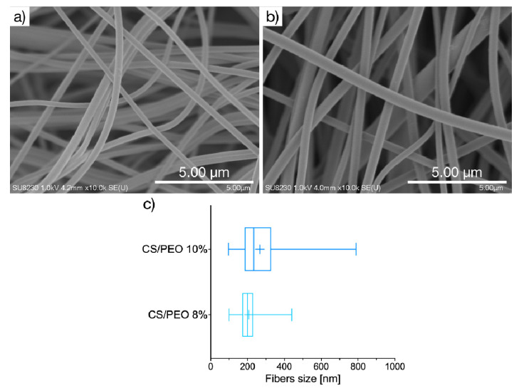
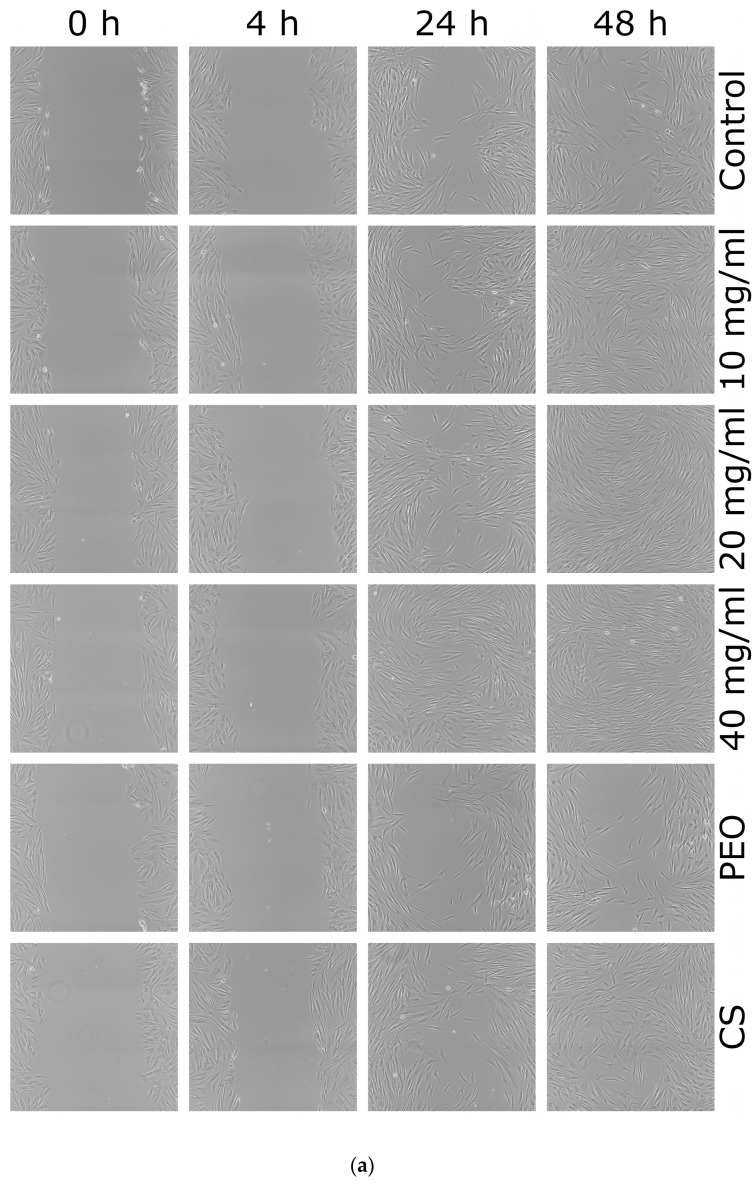
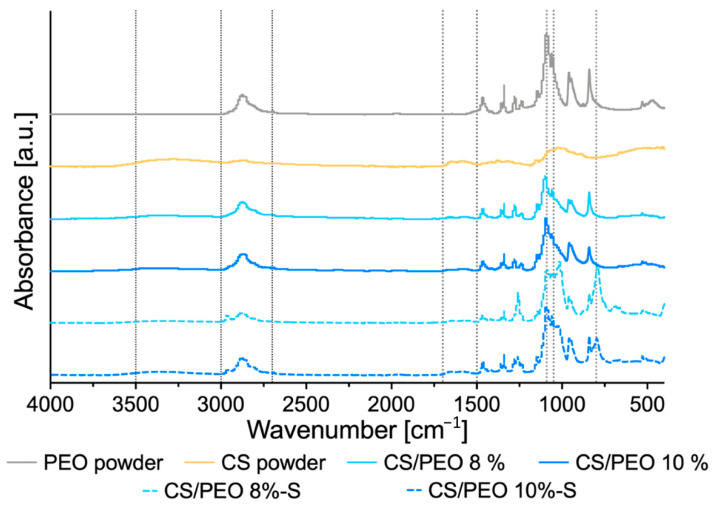
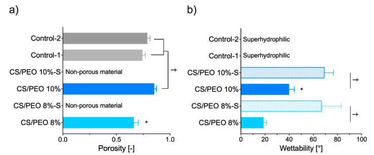
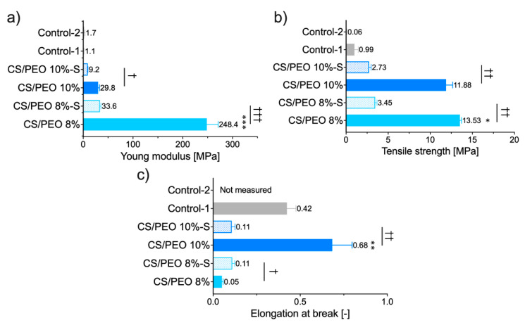
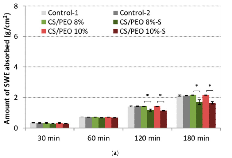
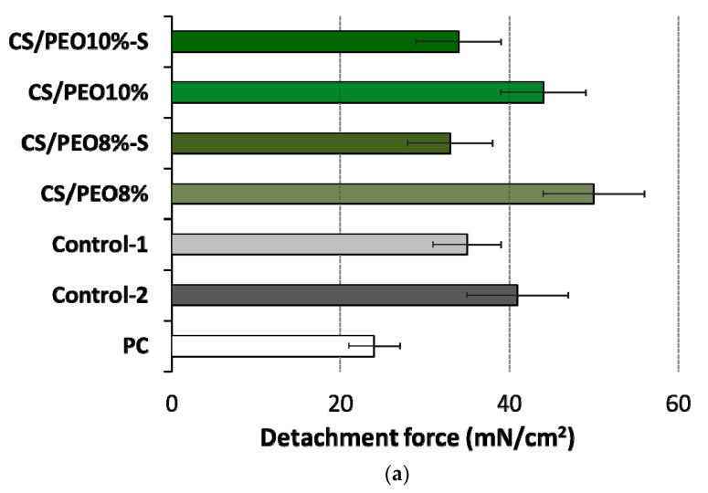
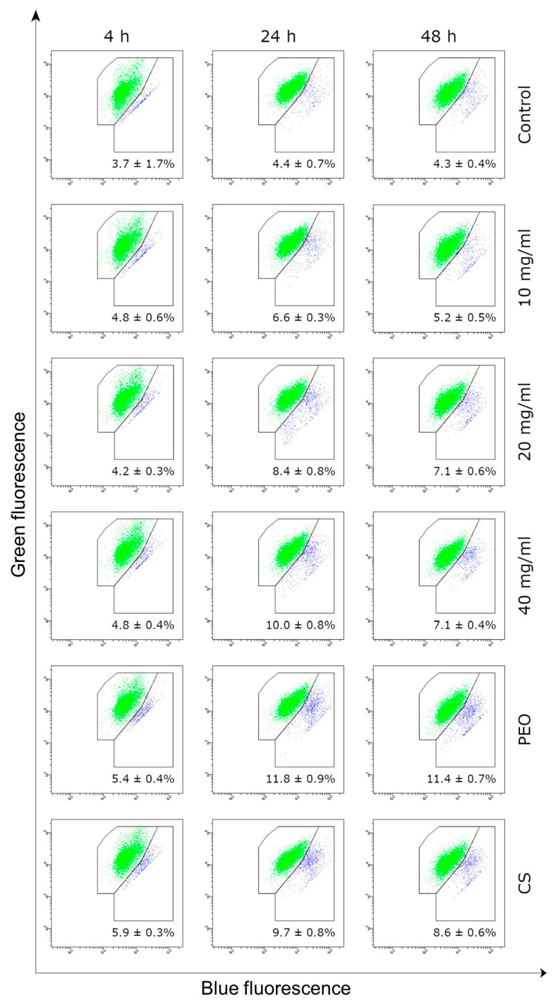
Chitosan (CS)/poly(ethylene oxide) (PEO)-based nanofiber mats have attracted particular attention as advanced materials for medical and pharmaceutical applications. In the scope of present studies, solution blow spinning was applied to produce nanofibers from PEO and CS and physicochemical and biopharmaceutical studies were carried out to investigate their potential as wound nanomaterial for skin healing and regeneration. Additional coating with hydrophobic poly(dimethylsiloxane) was applied to favor removal of nanofibers from the wound surface. Unmodified nanofibers displayed highly porous structure with the presence of uniform, randomly aligned nanofibers, in contrast to coated materials in which almost all the free spaces were filled in with poly(dimethylsiloxane). Infrared spectroscopy indicated that solution blow technique did not influence the molecular nature of native polymers. Obtained nanofibers exhibited sufficient wound exudate absorbency, which appears beneficial to moisturize the wound bed during the healing process. Formulations displayed greater tensile strength as compared to commercial hydrofiber-like dressing materials comprised of carboxymethylcellulose sodium or calcium alginate, which points toward their protective function against mechanical stress. Coating with hydrophobic poly(dimethylsiloxane) (applied to favor nanofiber removal from the wound surface) impacted porosity and decreased both mechanical properties and adherence to excised human skin, though the obtained values were comparable to those attained for commercial hydrofiber-like materials. In vitro cytotoxicity and irritancy studies showed biocompatibility and no skin irritant response of nanofibers in contact with a reconstituted three-dimensional human skin model, while scratch assay using human fibroblast cell line HDFa revealed the valuable potential of CS/PEO nanofibers to promote cell migration at an early stage of injury.
壳聚糖(CS)/聚环氧乙烷(PEO)基纳米纤维垫作为医学和制药应用的先进材料引起了特别关注。在本研究范围内,应用溶液吹纺法制备 PEO 和 CS 的纳米纤维,并进行物理化学和生物制药研究,以研究其作为伤口纳米材料促进皮肤愈合和再生的潜力。进一步用疏水性聚二甲基硅氧烷(PDMS)进行涂层,有利于从伤口表面去除纳米纤维。未修饰的纳米纤维具有高度多孔的结构,存在均匀的、随机排列的纳米纤维,而涂层材料中的几乎所有自由空间都被 PDMS 填充。红外光谱表明,溶液吹纺技术不会影响天然聚合物的分子性质。所得纳米纤维具有足够的伤口渗出液吸收能力,这有利于在愈合过程中保持伤口床的湿润。与由羧甲基纤维素钠或藻酸钠钙组成的商业水纤维状敷料材料相比,制剂的拉伸强度更大,这表明它们具有抵抗机械应力的保护功能。用疏水性 PDMS(有利于从伤口表面去除纳米纤维)进行涂层会影响孔隙率,并降低机械性能和对离体人皮肤的粘附力,尽管获得的数值与商业水纤维状材料相当。体外细胞毒性和刺激性研究表明,纳米纤维与重建的三维人皮肤模型接触时具有生物相容性,且无皮肤刺激性反应,而用人成纤维细胞系 HDFa 进行划痕试验表明,CS/PEO 纳米纤维具有促进细胞在受伤早期迁移的有价值的潜力。